More than a decade ago, Max Diem, professor of chemistry and chemical biology at Northeastern University, started thinking of a way to improve health care. “Most of medical diagnoses were done by pathologists or cytologists using methods developed 50 or 60 years ago,” he says. Instead of relying on what someone sees by eye, though, Diem wanted to add objectivity to the techniques. “If you could measure the chemical composition of a pixel of cell or tissue–say, a volume that is about 10 µm on a side and 2 µm deep–it could lead to reproducible, quantitative measurements,” he says. To pursue that idea, he turned to infrared spectroscopy.
Infrared (IR) light–wavelengths from about 2.5 to 25 µm, but varying some from one naming scheme to another–is longer than humanly visible wavelengths. In describing the benefits of using IR or near-IR for microscopy, Nicolas B. George, group marketing manager for light microscopes at Olympus America, says, “These longer wavelengths are relatively low energy, and their effect on living cells and tissue can be relatively benign when compared to shorter wavelengths of UV and visible light.” In addition, an infrared spectrum is inherently more sensitive than visible spectra, since it contains dozens of fingerprint bands to all major chemical constituents.
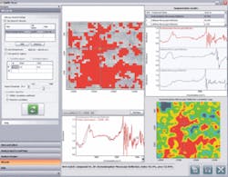
Inspired by IR’s beneficial features, Diem developed a system that can scan samples; he analyzes their data with chemometrics. “We can correlate the spectral information with standard histopathology or cytopathology,” Diem says. “We are training our method against classical methods of diagnosis.” Eventually, Diem hopes that his IR system of spectroscopy will make diagnoses on its own.
In the meantime, IR microscopy offers benefits that biologists can already use.
Defeating the diffraction limit
In any form of microscopy, biologists face two challenges: contrast and spatial resolution. “For 30 years,” says Shyamsunder Erramilli, professor of physics and biomedical engineering at Boston University, “people used visible microscopy and added stains or fluorescent labels for contrast. We get used to thinking of biological molecules being colored, like skin and blood, but the overwhelming majority of molecules are completely colorless.” Overcoming the colorless nature of biology with labels, however, is not always a good choice. “Stains can perturb the sample,” says Erramilli.
With mid-IR illumination–around 3 to 12 µm–many biological molecules vibrate. “They are playing music,” says Erramilli. “If you could see biological tissues or cells in mid-IR, the contrast would be amazing.” The trouble is that those wavelengths provide too little spatial resolution, at least for conventional microscopy. So, Erramilli uses near-field microscopy, which was invented–at least theoretically–in the mid-1920s by Irish scientist E.H. Synge. “In near-field microcopy, you illuminate an object through a tiny hole that is closer to the object than the wavelength of the light,” says Erramilli. “Then you move the hole a bit–in steps smaller than the wavelength of the light–and illuminate a different spot.”
To make this technique work better, Erramilli would like companies to build an IR microscope from the ground up. “Most IR microscopes can be tracked back to a visible-microscope platform,” he says. Consequently, he says, most of their parts are not optimized for IR microscopy. “I’m really disappointed in the source,” he says. “They’re still using glow bars, which is 40-year-old technology.” Instead, he uses a tunable laser, but his is worth about half a million dollars–hardly suitable for a production scope.
That price, though, pays off for Erramilli, because he can get below the diffraction limit with his system. “We are interested in finding lipids or proteins in membranes,” he says. “We could also look at the nucleus and quantify DNA.”
Mixing methods
Although biologists want to use a range of wavelengths to illuminate samples, they do not want a different microscope for every form of light. Instead, biologists want one microscope that handles a range of illumination wavelengths, but that takes some technological tweaks. “Much of the challenge involves providing chromatic correction throughout the range of observation,” says George of Olympus. In addition, the objectives must have higher numerical apertures to provide adequate resolution and light gathering.
“Olympus offers the UIS2 line of optical components and accessories, which are designed to meet these challenges,” George says. “The UIS2 objectives feature a dramatic improvement in near-IR transmission, along with improved transmission down to 340 nm.” For example, the U Plan S Apo “Super Apo” objectives, says George, “offer chromatic correction into the near-IR and hold specimen focus when switching between blue- and red-excited dyes.”
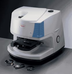
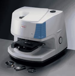
In some cases, though, companies also make microscopes with more-specific purposes in mind. For example, Thermo Fisher Scientific recently released its Nicolet iN10 MX infrared imaging microscope. Likewise, PerkinElmer recently released its Spotlight 400 mid- and near-infrared microscope and imaging system.Using such images, though, depends on more than optics. “The huge amount of data, which can be generated in minutes, has naturally placed additional burdens on the image-analysis software, and we have developed various hyperspectral image-analysis routines to enable information to be extracted from images much more efficiently,” says Sellors.
Beyond multiple wavelengths, a variety of modern imaging techniques rely on multiple photons, often to reduce the energy aimed at a living tissue. With such applications in mind, Olympus developed its Fluoview FV1000-MPE, which is a high-performance multiphoton system that works with IR. “Multiphoton excitation is ideal for live-cell imaging, because it allows deep penetration into the specimen with less photobleaching,” says George.
Overall, the expanding range of IR approaches gives biologists new views of nature. These views, however, extend beyond wavelengths of light, and might lengthen the span of life itself.
About the Author
Mike May
Contributing Editor, BioOptics World
Mike May writes about instrumentation design and application for BioOptics World. He earned his Ph.D. in neurobiology and behavior from Cornell University and is a member of Sigma Xi: The Scientific Research Society. He has written two books and scores of articles in the field of biomedicine.


