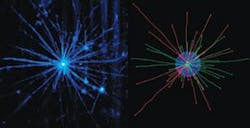
Scientists at the European Molecular Biology Laboratory (Heidelberg, Germany) have gained fresh insights into processes that occur inside biological cells by using a new form of microscopy that permits them to image samples in three dimensions. Their technique—laser light-sheet-based fluorescence microscopy (LSFM)—has provided the first dynamic images of microtubules, the networks of protein filaments that constitute major parts of cells’ skeletons (see figure).
“We were actually able to measure the mechanical properties of microtubules as a function of their state,” says team leader Ernst Stelzer.
Laser light-sheet-based fluorescence microscopy overcomes a major problem that users of conventional and confocal fluorescence microscopes often encounter: when a single plane is recorded, the microscopes cause phototoxic damage almost everywhere throughout the target sample. The LSFM technique avoids that difficulty by illuminating specimens along an entire plane with a light sheet and observing the emitted light orthogonally with a conventional fluorescence microscope.
“Simply observing causes negative effects,” Stelzer says. “An LSFM provides optical sectioning directly. It only excites the fluorophores in the illuminated plane and completely avoids photobleaching and other photo-induced damage outside the thin volume of interest.”
This feature makes LSFM particularly well suited for imaging sensitive biological objects over long periods of time. To facilitate the observation of microtubules, Stelzer’s team completely revised its procedures for preparing the cell samples.
“Instead of squeezing the asters [the fibrils that contain microtubules] between two hard, flat pieces of glass, we allowed them to develop in three dimensions,” Stelzer says. “This means their behavior is less restricted, and hence probably closer to physiological conditions.” Subsequent observation with the LFSM captured that behavior in full detail as it quickly recorded multiple slices of the cells samples.
Stelzer points to one highlight of the LFSM-assisted studies: biologists have known that microtubules shrink and expand. His group found that the components spend more time pausing— neither shrinking nor expanding—than shrinking. As a result of that and other findings, he and his colleagues have proposed an entirely new model for the behavior of microtubules, with considerably more parameters that have to be measured and taken into account. In fact, the development of the new microscope technology holds promise for other studies of cell biology.
“LSFMs are simple and much cheaper than confocal and two-photon microscopes, and they reduce photobleaching and phototoxicity dramatically,” Stelzer says. “I am absolutely sure that such instruments will be available all over the world within a few years.”
Imaging cellular components
In related news, another European team has developed a similar means of imaging different cellular components in their natural surroundings. Researchers at the University of Bordeaux in France have produced high-contrast images of mitochondria in living cells. The group’s technique, light-induced scattering around a nanoabsorber, uses a laser to heat up mitochondria and the regions around them. The increase in temperature induces a time-modulated variation of the refractive index around the mitochondria. When a probe beam interacts with that index profile, it produces a scattered field that is detected by a microscope objective.
“This method is highly original in microscopy because it is based on absorption and does not suffer from any background, even in the highly scattering autofluorescent environment of living cells,” says team member Laurent Cognet. “What’s more, the resolution we obtain is comparable to that of confocal microscopes.”
About the Author
Peter Gwynne
Freelance writer
Peter Gwynne is a freelance writer based in Massachusetts; e-mail: [email protected].
