PHOTOACOUSTIC MICROSCOPY: Isolating signal from photobleached molecules produces super-resolution image
More Biooptics World Articles |
INSIDE INSTRUMENTATION: Deep down and label-free: Bioimaging with photoacoustics |
Researchers at Washington University in St. Louis (MO) have turned a significant drawback of laser microscopy—photobleaching—into an equally significant advantage. "Previously when a molecule was prone to bleaching, researchers didn't want to use it because they couldn't get enough information from it," explained postdoctoral researcher Junjie Yao. "Now for us, that is good news."1
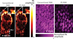
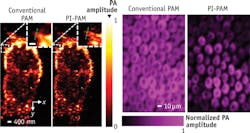
The center of a laser beam is so strong that it bleaches a specimen's midpoint, rendering it unable to produce information-packed signal. A second laser pulse probes the molecules in the boundary of the sample, and extracts a relatively weak signal from the bleached area. Yao and colleagues in the lab of photoacoustic microscopy pioneer Lihong V. Wang found that they could collect data from the center of a specimen illuminated by a 200-nm-diameter laser beam by subtracting this boundary area. What's left is a super-resolution image of the 80-nm-diameter center—what they call a photo imprint. The approach effectively shrinks the detection spot, allowing a view of subcellular structure, including features such as mitochondria and cell nuclei.
After each area of the sample is scanned, the researchers create an image. With previous photoacoustic microscopy imaging, the microspheres on the image were blurry. However, with the new photo-imprint photoacoustic microscopy, the result is clear and sharp.
Other scientists working in imaging could apply this method to their own research, Yao said.
1. J. Yao, L. Wang, C. Li, C. Zhang, and L.V. Wang., Phys. Rev. Lett., 112, 014302 (2014).

