BIOIMAGING/MICROSCOPY/NEUROSCIENCE: Low cost and advanced imaging apply beyond Neuroscience audience
Low cost was a theme at the 2013 Society for Neuroscience annual meeting (November 9–13, San Diego, CA), as vendors demonstrated their responses to the effect of funding cuts on researchers. But neuroscientists won't be the only ones to benefit from the exhibited products.
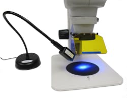
NIGHTSEA's (Bedford, MA) Stereo Microscope Fluorescence Adapter (SFA) system was an attention-getter, both for its price (from less than $1,000 to under $2,500) and for the story of its development by founder and president Charles Mazel. An add-on that enables existing lab-level stereomicroscopes to participate in the trend toward fluorescence that has been dominated by high-end stereomicroscopes, the expandable SFA offers four interchangeable excitation/emission combinations, with excitation wavelengths including violet, royal blue, cyan, and green.
Another offering for fluorescence microscopy, QImaging's (Surrey, BC, Canada) new optiMOS scientific CMOS (sCMOS) camera is, at $9,950, "the first product that delivers on sCMOS performance, yet is accessible within the constraints of typical fluorescence microscopy budgets," according to product manager Chris Ryan. An alternative to CCDs and capable of imaging cell dynamics, it promises fast frame rates (2.1 Mpixels at 100 frames/s), low noise, high resolution, and sensitivity to resolve low luminescence signals.
"An extremely cost-effective option" is how Nikon Instruments (Melville, NY) describes its LU-NA four-line solid-state laser system for microscopy. Like the LU-NB, which accommodates up to eight laser lines, LU-NA requires no calibration, guarantees permanent alignment, and ensures maximum power throughput—thus addressing the major limitations of traditional laser launches. In addition, LU-NB is particularly suited for Nikon's new Ti-LAPP system, which allows up to 32 imaging modalities to be combined into a single microscopy platform. In fact, multimodal imaging—and more capable imaging—were other themes in the exhibit hall.
More microscopy
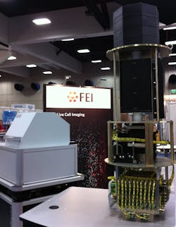
FEI Company (Hillsboro, OR) has come a long way (through a number of acquisitions) from its original focus on nonoptical microscopy: The company displayed an array of optical systems and supporting products, including the 5.5 version of its Amira 3D analysis software, which offers such functions as neuron tracing for neuroscientists. FEI's iMIC motorized light microscopy platform has demonstrated proficiency in combining different imaging modalities, and allowing easy switching between them and among applications—even within an experiment.1 And its CorrSight correlative microscopy offering aims to optimize the major steps of a workflow involving both light and electron modalities.The only other company addressing correlative microscopy to date is Zeiss (Thornwood, NY), which also showed a new module for its ELYRA P.1 super-resolution photoactivated localization microscopy (PALM) system: The module allows 3D imaging of endogenously expressed, photo-switchable fluorescent proteins in a range of 1.4 μm—and treats specimens with extra care to allow long-term observation. Zeiss also emphasized its light-sheet microscopy systems (see "The planar truth about light-sheet microscopy"), and Applied Scientific Instrumentation (ASI; Eugene, OR) offered "all of the necessary hardware for generating the light sheet from a laser, and for scanning the sample."
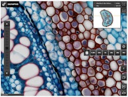
Olympus (Center Valley, PA) showed off its OlyVIA Mobile iPad App (a free download from iTunes), which provides access to your entire library of microscope imagery at various resolutions. Olympus focused its annual neuroimaging symposium on cellular-resolution functional brain imaging, with researchers from the Stanford University lab of optogenetics pioneer Karl Deisseroth.A new kid on the microscopy block, Vutara (Salt Lake City, UT) was on hand to discuss its SR-200, designed for acquiring and analyzing super-resolution images with "speed, simplicity, and stability." Its technique provides lateral resolution of 20 nm, axial resolution of 50 nm, up to 5 μm imaging depth (with z-stack acquisition), simultaneous two-color imaging in super-res mode (up to four colors in wide-field mode), and 3D particle tracking with ~10 nm precision.
Also new and cool
Among other new products on display was the Simoa single-molecule measurement system, a fluorescence-based diagnostic platform promising sensitivity 1000X greater than any other technology on the market. Maker Quanterix (Lexington, MA) delivered a poster describing a pilot study done with the Swedish Hockey League using Simoa to analyze a single drop of blood for biomarkers indicating brain injury—an alternative to spinal tap.
Dage-MTI (Michigan City, IN) emphasized simplicity (through USB connection, among other features) with both its real-time cameras: The newly designed IR-1000 for electrophysiology (with automatic control and edge enhancement) and its high-definition HD-210U with true 1920 × 1080 resolution, live 60 frames/s video, and both RGB and CMYK color.
Prior Scientific (Rockland, MA) previewed its ultra-quiet HLD117, "the most repeatable microscope stage available today" with 50 nm encoders. Kendall Research Systems (Cambridge, MA) talked about its forthcoming wireless optogenetics products. And Intelligent Imaging Innovations (3i; Denver, CO) offered "the only" commercially available spatial light modulator (SLM) designed for patterned and 3D point photomanipulation in optical microscopy.
Doric Lenses (Quebec City, QC, Canada) produced an optogenetics hardware catalog for the neuroscience audience, while Aplegen (Pleasanton, CA) promoted its Omega Lum G imaging system, "a versatile machine and quite powerful for the price," according to neurobiology professor Eric Norstrom of DePaul University.
Finally, Inscopix (Palo Alto, CA) discussed its strategy of selling only to selected customers; the company's Neuroscience Early Access Program (NEAP) invites labs to apply for the opportunity to work with nVista HD, "the first end-to-end solution for visualizing large-scale neural circuit dynamics in freely behaving animals." And TissueVision (Cambridge, MA) displayed TissueCyte 1000, which can image whole organs in four hours-and is being used by the Allen Institute for Brain Science for their open online Allen Mouse Brain Connectivity Atlas project: http://connectivity.brain-map.org.
Reference
1. J. Lin and A. D. Hoppe, Microsc. Microanal., 19, 350–359 (2013); doi:10.1017/S1431927612014420.

Barbara Gefvert | Editor-in-Chief, BioOptics World (2008-2020)
Barbara G. Gefvert has been a science and technology editor and writer since 1987, and served as editor in chief on multiple publications, including Sensors magazine for nearly a decade.


