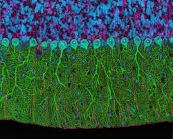
The annual Olympus BioScapes Digital Imaging Competition honors still and moving images of human, plant, and animal subjects as captured through light microscopes. Entrants can use any light microscope; entries are judged based on the science they depict, their beauty or impact, and the technical expertise involved in capturing them.
First Prize in the 2014 awards went to a team from the Howard Hughes Medical Institute Janelia Research Campus (Ashburn, VA) for their video clip depicting development of a fruit fly. In the fascinating video, a ball of cells turns into a fully developed larva that starts to crawl off screen. High-speed video like this allows researchers to follow the fate of individual embryonic cells. The movie was captured using a simultaneous, multi-view light-sheet microscope; its true potential is extracted by using complex mathematical models to learn the fate of every cell.
Second Prize (see figure) was given to Thomas Deerinck of the University of California San Diego's National Center for Microscopy and Imaging Research for a brilliant multiphoton image of a rat's cerebellum, captured at 300x. All award-winning entries are online at www.olympusbioscapes.com.
About the Author

Barbara Gefvert
Editor-in-Chief, BioOptics World (2008-2020)
Barbara G. Gefvert has been a science and technology editor and writer since 1987, and served as editor in chief on multiple publications, including Sensors magazine for nearly a decade.
