PRODUCT FOCUS: Microscope nanopositioning
Nanopositioning—which provides motion control at a fast rate increments of 1 nm or less over a range of several microns1—is a key enabler for such super-resolution microscopy techniques as photoactivated localization microscopy (PALM), spatially modulated illumination (SMI), and stimulated emission depletion (STED). A nanopositioner—usually a closed-loop control system—comprises a mechanical stage that consists of a movable component, inside a rigid frame, that can move in one or more of three translational axes (x, y, and z), as well as in angular focus feedback r axes such as rotation or tilt. Closed-loop control allows a user to provide inputs to the positioner and to monitor its parameters.2 I spoke with four different nanopositioning system makers about some recent innovations in this product category, and what they mean for life scientists.
Advantages in bio
Nanopositioners offer precision and repeatability of the adjustment paths, says Sebastian Reuter, public relations manager at Piezosystem Jena (Jena, Germany). Such accuracies can improve research results in advanced microscopy applications. Many companies are now modifying their optical microscopes to perform 3-D analysis, which requires piezo-grade precision and repeatability, he adds. John Zemek, executive director at Applied Scientific Instrumentation (ASI; Eugene, OR), notes that nanopositioners enable samples to be precisely moved to multiple locations during a time-lapse experiment and focus to be maintained during experiments, thereby yielding more accurate results.
Continuous motion with sub-nanometer precision and repeatability is key for high-resolution 3-D confocal imaging—and just isn't possible with motors or open-loop systems, among others, says Jenice Con Foo, marketing manager at Mad City Labs (Madison, WI). "There isn't much point in looking at features below the diffraction limit if you cannot maintain a stable reference frame," she says.
Scott Jordan, director, NanoAutomation Technologies, Physik Instrumente (PI; Auburn, MA), points out the importance of nanopositioning in imaging living cells. "The cell is a busy place with much more to look at than just enzymes, proteins, the nucleus, and the mitochondria," he says. He compares the cell to a cityscape, with a latticework of superhighways and feeder roads on which molecular motors carry payloads from place to place, while other nano-mechanical players assemble proteins, express genes, translate RNA, etc. So nanopositioning equipment works to bring the inner workings of cells together for proper imaging via several options, including long-travel, coarse-positioning sample stages with high stability; autofocus mechanisms capable of switching instantly to nanopositioning mode; and thin positioners that integrate into microscopes and mix with most imaging suite software.
Recent innovations
Zemek notes that high-resolution, linear-encoded servo motor stages with embedded piezos within the top plate of the stage allow movement with resolutions in the 5–10 nm range throughout the 120 × 100 mm x-y travel range of the stage. What's more, the embedded piezos for z positioning allow fast z-stacks to be taken with resolution in the 1–2 nm range for super-resolution microscopy applications.
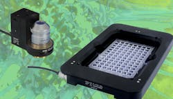
A Z-table is a piezo microscope stage that works in the z direction and has a standard k-frame aperture that can accommodate nearly all standard inserts, says Reuter. This stage allows the user to not only use common types of specimen tables, but also to work with various microscopes with different tables, he says (see Fig. 1).
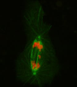
In establishing that stability plays a key role in microscopy, Mad City Labs has answered the call: Their Nano-Cyte 3-D active stabilization system enables single-molecule techniques in various cell biology applications, says Con Foo. The integrated system eliminates microscope drift and provides 3-D stabilization at the nanometer scale for days, she says.Feedback sensors-including linear encoders, laser interferometers, and custom systems-are critical in maintaining a constant sample position even as the thermal environment changes, which is key for life sciences work. An example of a custom system includes ASI's recently launched Continuous Reflective-Interface Sample Placement (CRISP) autofocus system, which provides focus feedback from the sample, explains Zemek (see Fig. 2). Able to be retrofitted to any microscope, the system allows researchers to maintain nanometer positioning for long-term experiments, he says, and maintain focus while scanning.
References
1. V. Coffey, Laser Focus World, 46, 5, 75–78 (2010).
2. http://www.npoint.com/resources.php#general1.

Lee Dubay | Managing Editor
Lee Dubay is managing editor for Laser Focus World. She is a seasoned editor and content manager with 20 years of experience in B2B media. She specializes in digital/print content management, as well as website analytics, SEO, and social media engagement best practices.

