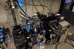
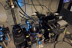
MICROSCOPY: New light-sheet microscope produces images in real time
Jan Huisken, research group leader of Max Planck, explained, "We exploited the spherical geometry of the embryos and the speed and sensitivity of the Zyla sCMOS camera [Andor Technology, Belfast, Ireland] to compute a radial maximum intensity projection of each individual embryo during image acquisition. An entire zebrafish embryo can now be instantaneously projected onto a 'world map' to visualize all endodermal cells and follow their fate. This reveals characteristic migration patterns and global tissue remodeling in the early endoderm and, by merging data from many samples, we have uncovered stereotypical patterns that are fundamental to embryo development."
The camera data were not saved at any point, so any 3D information not captured in the projection was lost. But the radial projection delivered immediate, pre-processed data for analysis and enabled experiments to be repeated rapidly. "This new technique will not eliminate the need for slow 3D imaging techniques in more complex shapes, but it is a highly effective strategy to streamline further analysis and increase throughput in many applications," he said.
1. B. Schmid et al., Nat. Comm., 4, 2207 (2013); doi:10.1038/ncomms3207.

