Photoacoustic microscopy images functions in the living brain quickly
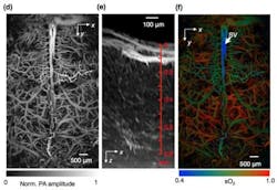
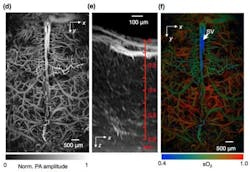
Using a photoacoustic imaging method, researchers at Washington University in St. Louis (WUSTL; Missouri) were able to see blood flow, blood oxygenation, oxygen metabolism, and other functions inside a living mouse brain at faster rates than ever before.
Related: Photoacoustic tomography is ready to revolutionize
The researchers used photoacoustic microscopy (PAM), a single-wavelength, pulse-width-based technique that was developed in the lab of Lihong Wang, Ph.D., the Gene K. Beare Professor of Biomedical Engineering in the School of Engineering & Applied Science, who led the work. The researchers were able to take images of blood oxygenation 50 times faster than their previous results using fast-scanning PAM; 100 times faster than their acoustic-resolution system; and more than 500 times faster than phosphorescence-lifetime-based two-photon microscopy (TPM).
Other existing methods, including functional magnetic resonance imaging (fMRI), TPM, and wide-field optical microscopy, have provided information about the structure, blood oxygenation, and flow dynamics of the mouse brain. However, those methods have speed and resolution limits, Wang says.
To make up for these limitations, Wang and his lab implemented fast-functional PAM, which allowed them to get high-resolution, high-speed images of a living mouse brain through an intact skull. This method achieved a lateral spatial resolution of five times finer than the lab's previous fast-scanning system; 25 times finer than its previous acoustic-resolution system; and more than 35 times finer than ultrasound-array-based photoacoustic computed tomography.
Most importantly, PAM allowed 3D blood oxygenation imaging with capillary-level resolution at a one-dimensional imaging rate of 100 kHz, or 10 µs.
"Using this new single-wavelength, pulse-width-based method, PAM is capable of high-speed imaging of the oxygen saturation of hemoglobin," Wang says. "In addition, we were able to map the mouse brain oxygenation vessel by vessel using this method."
"Much of what we have learned about human brain function in the past decade has been based on observing changes in blood flow using functional MRI," says Richard Conroy, Ph.D., program director for Optical Imaging at the National Institute of Biomedical Imaging and Bioengineering. "Wang's work dramatically increases both the spatial and temporal resolution of photoacoustic imaging, which now has the potential to reveal blood flow dynamics and oxygen metabolism at the level of individual cells. In the future, photoacoustic imaging could serve as an important complement to fMRI, leading to critical insights into brain function and disease development."
Concerned about the effects of the microscopy method on the living tissue, Wang and his team found that all red blood cells that were imaged were intact, and there was no damage to brain tissue.
"PAM is exquisitely sensitive to hemoglobin in the blood and to its color change due to oxygen binding," Wang says. "Without injecting any exogenous contrast agent, PAM allows us to quantify vessel by vessel all of the vital parameters about hemoglobin and to even compute the metabolic rate of oxygen. Given the importance of oxygen metabolism in basic biology and diseases such as diabetes and cancer, PAM is expected to find broad applications."
Full details of the work appear in the journal Nature Methods; for more information, please visit http://dx.doi.org/10.1038/nmeth.3336.
-----
Follow us on Twitter, 'like' us on Facebook, connect with us on Google+, and join our group on LinkedIn

