Intravital microscopy can study virus responses
Research at the San Raffaele Scientific Institute (Milan, Italy) applies intravital microscopy to the study of host viruses and associated immune responses.
Related: Super-resolution microscopy helps quantify viral DNA
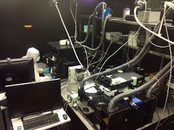
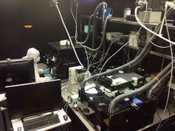
Led by Matteo Iannacone, MD, Ph.D., the group leader in the Division of Immunology at the San Raffaele Scientific Institute, the work seeks to dissect the complex dynamics of host virus interactions, with a particular focus on the development and function of adaptive immune responses. Since it is still beyond the reach of sophisticated in vitro methodology to simulate the complex interplay of physical, cellular, biochemical, and other factors that influence cell behavior in microvessels and interstitial tissues, the group uses intravital microscopy. This technique is complemented by more traditional molecular, cellular, and histological approaches, thus characterizing host virus interactions at the molecular, single-cell, and whole-animal level. Iannacone says that the group couples a state-of-the-art multiphoton microscope to enable in vivo studies. They also use confocal immunofluorescence histology and correlative light electron microscopy (CLEM), he says.
Iannacone, in a collaboration with the Department of Physics at the University of Milano-Bicocca, is developing a new method where he will use multiphoton microscopy to map blood flow velocity. In vivo microscopy allows mapping of blood flow velocity at a high spatial and time resolution in the complex network of vessels constituting the hepatic microcirculation while simultaneously maintaining the morphologic information related to the dynamic parameters of vessels and immune cells. This considers the potential of various novel imaging techniques to show how CD8+ T-cells move through the liver and how this may impact on the pathogenesis of hepatitis B virus (HBV).
Full details of the work appear in the journal Cellular & Molecular Immunology; for more information, please visit http://dx.doi.org/10.1038/cmi.2014.78.
-----
Follow us on Twitter, 'like' us on Facebook, connect with us on Google+, and join our group on LinkedIn

