Low-light microscopy technique avoids damage to light-sensitive specimens
Seeking to help clarify low-light microscopy images while avoiding damage to delicate specimens, researchers at Stanford University have developed a technique that could make it possible to view proteins and living cells in greater clarity.
Related: New bioimaging technique offers clear view of nervous system
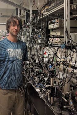
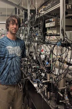
In optical microscopy, individual photons strike a detector to make the image. The researchers have found they get better results if each photon interacts with the sample multiple times, even in low light. To implement this in a microscope, instead of sending light through a specimen and then directly capturing the resulting image, the research team repeatedly reflects the image back onto the specimen. Study co-author Brannon Klopfer, a graduate student in Stanford professor Mark Kasevich's group at Stanford, explains that following the image taken of the specimen, you then illuminate it with an image of itself that again is sent back to illuminate the sample, leading to contrast enhancement.
The research team's technique, which they call multi-pass microscopy, is not the only approach to overcoming the shot-noise limit. Another technique, called quantum microscopy, uses entangled photons to achieve the same result, but it is more challenging to carry out. The ability of entangled photons to give information about each other means that quantum microscopy can produce higher-quality images compared to standard microscopy. At present, multi-pass microscopy has the potential to create comparably enhanced results with the added benefit of requiring less arduous preparation than quantum microscopy.
"The advantage you gain when entangling two photons is what we gain when we go through the sample twice," says Thomas Juffmann, co-author of the research and a postdoctoral research fellow in the Kasevich research group. "Currently, it is technologically easier to make a photon pass through a sample 10 times than to create a state in which 10 photons are entangled with each other."
Multi-pass microscopy could boost more than just low-light microscopy because it acts as a general signal-enhancing technique. The method can increase the sensitivity of various microscopy techniques, so long as a source of image noise doesn't build up with the recycling of photons.
At present, multi-passing is restricted to optical microscopes, but the research team is also working on multi-pass electron microscopy, where damage prevents the atomic-scale imaging of single proteins or DNA. Recycling the electrons in electron microscopy would improve image quality just as the recycling of photons does in the optical microscopes.
Full details of the work appear in the journal Nature Communications; for more information, please visit http://dx.doi.org/10.1038/ncomms12858.

