Ultrathin, flat microscope enables rapid melanoma detection
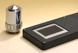
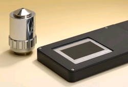
Researchers at the Fraunhofer Institute for Applied Optics and Precision Engineering (Jena, Germany) have designed a flat, lightweight microscope able to examine to a resolution of 5 µmâenabling it to make a single measurement and record across a broad imaging area. The initial purpose of the microscope is to enable melanoma detection on a section of a patient's skin in less than one second.
The microscope features a 5.3 mm optical length, making it flat and easy to use as a method of rapid detection. Tiny imaging channels, with lots of tiny lenses arrayed alongside one another, allow the microscope to record a tiny segment of the object at the same size for a 1:1 image, doing so channel by channel. Each segment measures roughly 300 x 300 µm² in size and fits seamlessly alongside the neighboring segment. Then, a computer program assembles all of the segments to generate the overall picture.
The imaging system consists of three glass plates with the tiny lenses applied to them, both on top and beneath. These three glass plates are then stacked on top of one another. Each channel also contains two achromatic lenses, so the light passes through a total of eight lenses. Several steps are involved in applying the lenses to glass substratesâthe scientists first coat a glass plate with photoresistant emulsion and expose this to UV light through a mask. The portions exposed to the light become hardened. If the plate is then placed in a special solution, all that remains on the surface are lots of tiny cylinders of photoresist; the rest of the coating dissolves away. Then, the researchers heat the glass plate: the cylinders melt down, leaving spherical lenses. Working from this master tool, the researchers then generate an inverse tool that they use as a die. A die like this can then be used to launch mass production of the lenses.
Researchers have already produced a first prototype and have showcased it at the LASER World of PHOTONICS 2011 trade fair in Munich (see http://bcove.me/wt0uzrsr). It will be at least another one to two years before the device can go into series production, according to the researchers.
-----
Posted by Lee Mather
Follow us on Twitter, 'like' us on Facebook, and join our group on LinkedIn
Follow OptoIQ on your iPhone; download the free app here.
Subscribe now to BioOptics World magazine; it's free!

