Ultrafast lasers advance deep-tissue imaging
Since the first landmark two-photon microscopy images in 1990, a wide range of nonlinear imaging methods that utilize ultrafast lasers have evolved, including three-photon fluorescence excitation, second- and third-harmonic generation (THG), coherent anti-Stokes Raman spectroscopy (CARS), and other types of nonlinear optical effects.1
Two advantages of multiphoton microscopy are its inherent ability to provide high-resolution three-dimensional images at significant depths and to do this in live tissue with minimal sample damage. Until recently, however, most multiphoton images have been taken near the sample surface with a typical depth of less than 500 µm. But compelling questions in fields such as embryonic morphogenesis, neurobiology, and angiogenesis can only be answered by probing farther into these live tissue samples, even as deep as 1 mm. This capability requires refining multiphoton excitation (MPE) tools.
As MPE techniques peer deeper into tissue, the signal-to-noise ratio steadily deteriorates. This is caused partly by scatter and absorption of the laser light, but is more critically due to scatter and absorption of the weak return signal that forms the images. From the laser perspective, there are three ways to boost the signal and thereby create brighter images in deep samples: use higher-power, shorter pulses, and/or longer excitation wavelengths. By definition, all multiphoton techniques have a high-order dependence on peak laser power; higher average power and shorter laser pulses both translate directly into higher peak powers. The use of longer excitation wavelengths lowers both scatter and absorption by the sample, which are the main causes of signal loss in deeper images. (Of course, these same “bright image” advantages also benefit applications involving shallower images, where they enable shorter data-acquisition times and hence minimized sample damage.)
Ti:sapphire has proved to be the key material enabling the development of user-friendly ultrafast lasers, including tunable one-box MPE lasers. However, compared to some other laser crystals, Ti:sapphire is a low-gain material. The only way to increase the output of a Ti:sapphire laser is to increase the pump power. For this reason, Coherent extended its Verdi integrated 532 nm pump lasers to produce output powers as high as 18 W, which results in ultrafast output as high as 3.5 W.
At the sample
There are two ways to deliver shorter pulses at the sample. The simplest method is to match the output of the laser to the group-velocity-dispersion (GVD) characteristics of the microscope. If the initial laser pulse is very long, it will be very long at the sample even though it does not undergo much additional GVD in the microscope optics. If the initial pulse is very short, it will undergo a considerable amount of GVD in the microscope. Therefore, there is an optimum input pulse duration. Coherent’s Chameleon lasers produce an output pulsewidth around 140 fs, which matches the GVD characteristics of most commercial microscopes and results in a pulsewidth at the sample of around 200 fs (see Fig. 1). For even shorter pulses, however, a turnkey accessory adds an adjustable amount of negative GVD, which can be set by the user to actually precompensate for the GVD of the microscope and any other beam-delivery optics. This enables pulses as short as 150 fs at the sample focus.
Achieving a longer wavelength range from a tunable Ti:sapphire laser requires a two-pronged approach: increase the optical gain and lower the cavity loss. The Ti:sapphire gain curve falls away beyond 1000 nm, limiting many commercial lasers to a long-wavelength limit around 1040 nm or less. The long-wavelength gain of a Ti:sapphire laser can be increased by using higher pump power, up to 18 W. In addition, redesigned broadband mirrors with ultralow scatter have lowered overall cavity losses. With this dual approach, the latest lasers can now routinely deliver continuously tunable output all the way to 1080 nm. Moreover, the addition of an OPO accessory extends the continuous tuning range as far as 1.5 µm.
In the specific case of MPE fluorescence microscopy, deeper imaging is also benefiting from the development of new fluorophores that can be used at these longer wavelengths. A standout example is the fluorescent protein mCherry which can be expressed in trangenic target cells and subcellular structures in a similar manner to the long-established GFP.
Probing angiogenesis
Angiogenesis is an area that can directly benefit from these deeper imaging tools. Mary Dickinson and coworkers at Baylor College of Medicine (Houston, TX) are using long-wavelength excitation and mCherry for this purpose. Systemic circulation is vital to normal cellular physiology and many factors can cause vessel growth and remodeling, including exercise, injury, diseases such as diabetes and stroke, as well as in the formation of tumors. Studying vessel and circulation dynamics deep within tissues in the body is essential to understanding the complexities of angiogenesis and the progression of disease.
To characterize cell and tissue dynamics during the formation of the cardiovascular system and in adult mice, Dickinson’s team generated a novel transgenic mouse line, Tg(Flk1::myr-mCherry), in which endothelial cell membranes are brightly labeled with mCherry. These Tg(Flk1::myr-mCherry) mice are viable, fertile, and do not exhibit any developmental abnormalities. High levels of mCherry are expressed in the embryonic endothelium and endocardium, and expression is retained in small capillaries in adult animals.2
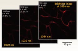
These researchers are studying angiogenesis in vivo with two-photon excitation of mCherry using excitation wavelengths beyond 1050 nm. Long-wavelength excitation is preferred for two reasons. First, they have found that the two-photon excitation efficiency is still increasing at 1060 nm (their modulator optics precluded data beyond this point). Therefore, longer wavelengths are more appropriate for the excitation peak of this fluorochrome (see Fig. 2). Second, the inherently reduced scatter and absorption at longer wavelengths improve both the delivery of the excitation light through the tissue, as well as aid in the recovery of emission signal, thereby increasing the sensitivity in scattering tissue. They believe that perhaps even greater improvements in signal detection can be made using even longer excitation wavelengths, even better than can be achieved using an OPO.“Currently, long-wavelength MPE is the only way to image these deep vessels at submicron resolution,” Dickinson says. “We use optical coherence tomography for other deeper studies, but this is limited to a spatial resolution of only 5 to 10 µm.”
Multiplex CARS
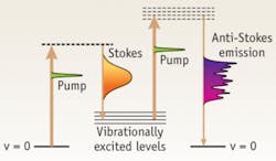
There is growing interest in using microscopy to probe structure and function without resorting to the use of stains, fluorophores, and fluorescent proteins (see Inside Instrumentation). Coherent anti-Stokes Raman spectroscopy (CARS) is a tool that has been shown to meet this need. First demonstrated by Sunney Xie and coworkers at Harvard University, CARS imaging is based on a so-called four-wave mixing technique using lasers at two wavelengths. A pump pulse, together with a Stokes pulse, excites the molecule to a vibrational state that is immediately probed by the pump pulse to generate the CARS signal at the anti-Stokes frequency. In conventional CARS imaging the frequency difference between the (fixed) pump and the (tunable) Stokes picosecond pulses is tuned to excite a single molecular Raman spectral resonance characteristic of a specific chemical of interest. Moving the sample relative to the focused laser waist, or vice versa, builds up an image that maps the distribution of that specific molecular type.
Multiplex CARS, as first demonstrated by Michiel Müller and coworkers at the Swammerdam Institute for Life Sciences, University of Amsterdam (Amsterdam, The Netherlands), steers this concept in a new direction. Here, the Stokes pulse is a broadband pulse and the resultant signal contains all the Raman peaks over a broad (~1500 cm–1) spectral region (see Fig. 3). The anti-Stokes emission is dispersed in a simple grating spectrometer and detected using a cooled linear CCD array. Each single multiplex CARS image thus contains information about the concentration and distribution of potentially every Raman active molecule and is even sensitive to subtle differences in local chemical environment.With current Ti:sapphire laser technology, there are actually several ways to generate multiplexed CARS images. One method uses a narrowband (picosecond) oscillator as the pump and a very broadband (femtosecond) laser oscillator as the Stokes. (A narrowband pump is critical to obtaining resolved spectral peaks.) Another uses a narrowband pump and a broadband supercontinuum as the Stokes, generated by coupling an ultrafast laser into a photonic-crystal fiber. The most elegant method uses just the output of a single broadband laser making optical alignment much simpler than the other methods that use two beams.
To meet this need for very broadband pulses, laser manufacturers have developed one-box, turnkey, ultrafast lasers with greater bandwidth than ever before. For example, the Coherent Micra laser oscillator has a bandwidth of more than 100 nm, which translates into more than 1500 cm–1 of Raman spectrum for each image pixel. This performance has been achieved by use of special cavity mirrors whose reflectivity is flat over a wide spectral range and whose GVD is linear over this range. These characteristics enable a prism-pair compensator in the laser cavity to correct for the GVD and deliver near-transform limited pulses.
As commonly happens with new laser technology, researchers often find other uses and applications that are enabled by the improved output characteristics. In this case, some biologists are taking advantage of the broad bandwidth of these new oscillators and using an optional pulse compressor to achieve pulses as short as 10 fs. They are investigating if and how such a short pulse impacts the relationship between image quality and sample damage in live tissue for THG and MPE fluorescence methods.
What can multiplex CARS do? A team led by Hervé Rigneault at the Institut Fresnel-Mosaic laboratory, CNRS/Paul Cezanne University (Marseille, France) has been developing CARS and multiplex CARS imaging systems for collaborative studies with biologists, including researchers from the Center of Immunology of Marseille Luminy (Marseille, France).3
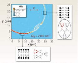
“We use a Coherent Micra laser to enable the single broadband laser method because the ease of optical alignment of a single beam means we can focus on our science, rather than optimizing the optical setup,” Rigneault explains. Specifically, they use a pulse shaper to retard the phase (by π) for a narrow spectral window as proposed by the team of Y. Silberberg at the Weizmann Institute of Science (Rehovot, Israel).4 The intensity variation due to the weak resonant signal is amplified by heterodyne detection with the strong nonresonant background, which acts as a local oscillator.The team is currently performing multiplex CARS with rotating linearly polarized laser beams. “With crystalline samples, it is well known that the CARS signal intensity is very dependent on the angle between the Raman transition dipole and the electric vector of the laser beam(s),” says Rigneault (see Fig. 4). “We are extending this concept to study model, and eventually real, membranes, to map the alignment of molecules within a membrane.”
REFERENCES
1. W. Denk, J.H. Strickler, and W.W. Webb, Science 248, 73 (1990).
2. I.V. Larina et al., Submitted for publication.
3. N. Djaker et al., Nuclear Instr. and Methods in Phys. Res. A 571, 177 (2007).
4. D. Oron, N. Dudovich, D. Yelin, and Y. Silberberg, Phys. Rev. Lett. 88(6), 063004 (2002).
Marco Arrigoni
Marco Arrigoni is vice president of marketing at Light Conversion (Vilnius, Lithuania). He previously served as director of marketing at Coherent (Santa Clara, CA) from 2007 through 2023.
David Armstrong | Product Manager, Coherent
David Armstrong is a Product Manager at Coherent (Santa Clara, CA).


