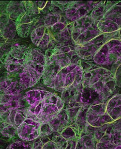
Two-photon microscopy method images oscillating mitochondria in rats
A two-photon microscopy method has enabled scientists at the National Institutes of Health's National Institute of Dental and Craniofacial Research (NIDCR; Bethesda, MD) to image mitochondria oscillating in a live animal—in this case, the salivary glands of laboratory rats. The research team's study shows that the oscillations occur spontaneously and often in the rodent cells, leading them to believe that the oscillations almost surely also occur in human cells.
Related: Two-photon microscopy method images neuronal activity across the entire brain
"The movements could last from tens of seconds to minutes, which was far longer and frequently at a faster tempo than observed previously in cell culture," says Roberto Weigert, Ph.D., an NIDCR scientist and senior author on the study. The mitochondria also appear to synchronize their movements not only in an individual cell but, quite unexpectedly, into a linked network of oscillators vibrating throughout the tissue.
The mitochondrion is of one of several distinct compartments, or organelles, in the cell cytoplasm. They generate a continuous supply of the molecule ATP that serve as the cell's main source of energy to power the heart to beat, muscles to stretch, and virtually every movement that the body makes. To keep cells fully charged, mitochondria operate four biochemical production lines that coalesce with oxygen molecules from normal respiration to produce ATP. One of these production lines starts with processing the molecule nicotinamide adenine dinucleotide (NADH). Weigert and colleagues recognized that they could use their intravital two-photon microscopy method to visualize NADH as it naturally emits electrons as part of the ATP production process.
Intravital two-photon microscopy, until recently, had been too powerful to use in live animals. "Animals breathe, their hearts beat, and their appendages twitch," says Weigert. "The combined effect under very high magnification is like watching a 6.0 earthquake. Everything shakes and blurs out of focus. We have developed approaches to better stabilize our organ of interest and minimize the motion artifacts. At this point, it is just a matter of generating more powerful optics to visualize the chemistry of life that really unfolds in the body, not under artificial laboratory conditions that stress cells and likely modify their behavior."
The powerful optics allowed the scientists to visualize the oscillations in their native milieu and to puzzle over their cause. Based on a series of subsequent experiments and observations, the researchers discovered that the oscillations are linked to the production of reactive oxygen species, a chemically interactive byproduct of making ATP. This finding suggests that the oscillations likely are not inherent to mitochondria, but a response to conditions in their environment.
"These findings emphasize how important it is scientifically to study biology on its own terms, not under artificial laboratory conditions," says Natalie Porat-Shliom, an NIDCR scientist and lead author on the paper. "We saw things in live animals that you don't see in cell culture. The reasons, in this case, very well may be that the mitochondria continue to receive an influx of signals from the blood vessels, the nervous system, and their surrounding environment. The entire system can't be reassembled in cell culture."
Porat-Shliom notes that these findings should be of broad interest scientifically in framing studies of mitochondria, and may have future clinical implications. An estimated 2 million Americans have mitochondrial disease, an energy-depleting failure of mitochondria to function properly, which can have disabling effects on the brain, heart, kidneys, and other body systems. Many scientists also suspect that as mitochondria become better understood, they likely will be understood to play a more prominent role in human health and disease.
Full details of the work appear in the journal Cell Reports; for more information, please visit http://dx.doi.org/10.1016/j.celrep.2014.09.022.
