Better results from ultrafast nonlinear microscopy
Precompensation for high-order chromatic dispersion makes possible the use of pulses as short as 10 fs for nonlinear optical microscopy. The resulting signal-to-noise increase can enable better-quality depth-resolved imaging.
By Marcos Dantus, Dmitry Pestov, and Yair Andegeko
Because of their inherent three-dimensional (3-D) sectioning capabilities, nonlinear optical microscopy methods such as two-photon excitation fluorescence1 (TPEF) and second-harmonic generation (SHG) are gaining popularity. With these methods, the signal is generated within a narrow region near the focus of the microscope objective, and it is well separated spectrally from the laser’s excitation wavelength. Photodamage, if any, is also spatially confined to the focal volume.
The laser parameter that makes these nonlinear methods possible is peak intensity, defined as energy per pulse divided by the area at the focus and the laser pulse duration. While the average laser power for nonlinear microscopy imaging is typically 1 to 10 mW, the peak intensity at the focus is as high as 1011 to 1012 W/cm2. These extremely intense fields cause molecules to absorb two or more photons at a time or induce a nonlinear polarization at the sample that gives rise to the second harmonic of the incident frequency.
The Dantus Research Group at Michigan State University’s Department of Chemistry has undertaken work to improve signal-to-noise ratio in nonlinear microscopy using ultrashort (≤15 fs) laser pulses. The rationale for using shorter pulses is a direct consequence of the fact that the peak intensity is inversely proportional to pulse duration. The number of TPEF or SHG photons is proportional to the square of the peak intensity times the pulse duration; therefore, the net signal is inversely proportional to the pulse duration at the focal plane.
Overcoming limitations
Laser availability has not been an issue; while most lasers used now for nonlinear microscopy have pulse durations of approximately 150 fs, a number of vendors offer ultrashort-pulsed lasers at prices that are comparable to the longer-duration-pulse systems. The challenge has been to compensate for the pulse broadening that occurs because of dispersion introduced primarily by the microscope objective.
FIGURE 2. TPEF and SHG images of an unstained murine bone specimen were obtained with fully compensated ultrashort laser pulses using MIIPS. The signal intensity for transform-limited pulses was sevenfold greater than the SHG signal acquired when only second-order dispersion correction via a prism-pair compressor was used. The pulse duration at the objective focus is 15 fs. Every 512 × 512-pixel image is averaged over 30 frames. Images are 150 × 150 µm.
The drive toward high spatial resolution and efficient signal collection has motivated the use of high-numerical-aperture microscope objectives. But high-numerical-aperture objectives have a drawback: those that are corrected for chromatic aberrations have the highest chromatic dispersion. As an ultrashort pulse is transmitted by one of these objectives, it experiences time broadening because different wavelengths take different amounts of time to travel through.
The key parameter for precompensation is the spectral bandwidth (full width at half maximum) of the laser. Since the earliest reports on TPEF microscopy, it was clear that precompensating for chromatic dispersion would be beneficial. Unfortunately, while correcting for second-order dispersion is quite simple, correcting for higher-order dispersion is much more complicated.
Improving TPEF and SHG efficiency
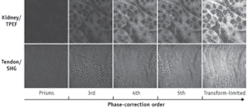
We looked at the signal intensity for both TPEF and SHG imaging as a function of precompensation for different orders of chromatic dispersion (see Fig. 1). From the acquired data it became clear that for pulses as short as 15 fs (FWHM bandwidth approximately 90 nm), correction of high-order dispersion is essential.2, 3 To obtain the data, we used a pulse shaper that allows manipulation of the phase of the laser pulses. In particular, we used the method known as MIIPS (multiphoton intrapulse interference phase scan) to measure and to precompensate the pulse dispersion.4
The results indicate the significant contribution of higher-order phase distortions. While the origin of those distortions is unclear, we believe they arise in part from the laser itself. The dielectric coatings used for generating ultrashort pulses may cause these types of distortions, too, especially at the edges of the spectrum. And the use of high-index glass for the microscope objective and the use of multiple antireflection coatings for the microscope objective may also produce significant high-order distortion.
FIGURE 3. Spectrally resolved SHG-TPEF images of murine footpad bone expressing green fluorescent protein were taken with ultrashort transform-limited laser pulses of 15 fs duration at the objective focus, after compensation via MIIPS. The SHG and TPEF 512 × 512-pixel images were acquired simultaneously in 60 seconds via 16-channel time-correlated single-photon counter. The average laser power on the sample was approximately 10 mW. Images are 150 × 150 µm.
Fortunately, all these issues can be addressed by accurate measurement of the phase at the focal plane and direct precompensation, both of which can be accomplished in approximately 25 seconds with the use of MIIPS.5
The ability to deliver pulses shorter than 15 fs results in greater amounts of signal per given pulse energy. This capability can be used to reach greater depth of penetration for depth-resolved imaging. In one instance, we used 15 fs pulses to image bone tissue at a depth exceeding 100 µm (see Fig. 2). In all cases, the use of longer pulses or only second-order precompensation resulted in approximately one order of magnitude less signal.
To illustrate how TPEF and SHG signals can be easily separated to distinguish different tissues, we imaged fluorescently labeled mouse footpad bone tissue at a depth of 50 µm, with all its living cells expressing green fluorescent protein (see Fig. 3). The bone tissue was mapped out by the SHG signal while the cells and blood vessels were visualized via TPEF imaging. TPEF and SHG separation was achieved spectrally using a multichannel time-correlated
Current and future benefits
These demonstrations confirm that the availability of ultrafast lasers, together with the technology for precompensating high-order chromatic dispersion at the focal plane, opens the possibility of using pulses as short as 10 fs for nonlinear optical microscopy. These ultrashort pulses provide a direct increase in signal-to-noise ratio that can be important for depth-resolved imaging. Similarly, the increased efficiency of the ultrashort pulses allows the use of considerably less laser power, which has been shown to reduce photobleaching.6
Our group is now exploring further methods–enabled by pulse shaping–for reducing photobleaching and photodamage in living cells. ‹‹
REFERENCES
- W. R. Zipfel et al, Nature Biotech. 21, p. 1368 (2003).
- P. Xi et al, J. Biomed. Optics 14, 014002 (2009).
- Y. Andegeko et al, SPIE Proc. Photonics West 2009.
- B. W. Xu et al, J. Opt. Soc. America B–Opt. Phys. 23, p. 750 (2006).
- Demonstrated at Photonics West 2009 by BioPhotonic Solutions (Okemos, MI)
- P. Xi et al, Optics Communications 281, p. 1841 (2008).
MARCOS DANTUS is professor, DMITRY PESTOV and YAIR ANDEGEKO are research associates, and is research associate in the Department of Chemistry, Michigan State University, East Lansing, MI; www2.chemistry.msu.edu/faculty/dantus/. Contact Dantus at [email protected].


