OPTICAL COHERENCE TOMOGRAPHY/PORTABLE INSTRUMENTATION: Portable OCT for point-of-care testing
Changho Chong, Daisuke Ogawa and Jonathan Evans
Point-of-care testing can provide continuous patient care away from central clinical facilities, and implies improvements in quality of care and life. The goal of this approach is to enable immediate testing and results so that clinical decisions can be made quickly and applied for better effect. To address this need and expand their locations of practice, however, clinicians require portable, compact, and robust systems.
An optical coherence tomography (OCT) system fitting this description promises to enable earlier detection and diagnosis of disease. Today, most commercially available OCT systems consist of a large desktop PC and a large probe head, and are either dedicated for specific imaging applications in ophthalmology and cardiovascular procedures, or are being used to study OCT itself. Researchers in OCT have applied the technique to a wide range of fields; however, outside of the lab, its use is still restricted. To encourage the wider adoption of the OCT technique, equipment must be more convenient and practical.
To address such point-of-care applications as home, remote health care, and emergency use, an imaging system should ideally be portable, versatile, reliable in its design, and easy to use. In fact, maintenance-free, robust equipment is required for doctors who use it on a daily or near-daily basis. Advances in hardware and novel techniques—including the recent implementation of cross polarization-sensitive detection, as well as usability through software—are enabling the realization of this vision.
Cross-polarization
One innovation adding to the usability of OCT is the technique of cross-polarization imaging, which adds stability and enhancement to the image quality.
Conventional OCT systems are based on polarization-insensitive detection techniques. Typically, a sample’s top surface exposed to the air has a strong specular reflection that attenuates the sub-surface signal of interest (such as that from a lesion). The standard method of reducing surface reflection and thereby enhancing imaging below the surface involves applying refractive index-matching gel on top of sample or immersing the sample in water. Not only are these approaches inconvenient for practical in-vivo situations, but they also serve to lower the value of the noninvasive nature of OCT. Cross-polarization overcomes this issue by selectively detecting only light of orthogonal polarization, with respect to the input light polarization. This type of probe can typically achieve a polarization extinction ratio greater than 30 dB.
Use of cross-polarization is especially effective in dental applications where the tooth surface is a strong reflector that interferes with a dentist’s wish to identify tooth decay or caries as early as possible. Clinicians at the National Center for Geriatrics and Gerontology implemented cross-polarization SS-OCT in Japan for the first time.1 Their work proves that reduction of surface reflection yields a clear image of the deep lesion; also, enhanced contrast allows for more accurate caries diagnosis and monitoring of re-mineralization processes (see Fig. 1). The advantages of cross-polarization can also be applied to other diagnostics such as oncology, where malignancy progresses from the subsurface of the organs.
Making a swept-source system compact
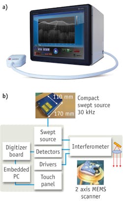
In addition to cross-polarization, several other key components—a swept light source, balanced detector, special digitizer board and CPU, and a small probe head—are combined in a compact (25 lb, <12 in. cube with touch-screen interface) design to provide an example of point-of-care functionality (see Fig. 2).The swept source is based on external cavity laser with a polygon scanner and diffraction grating filter.2 The compact polygon scanner, widely used in commercial laser printers, adds robustness and reliability. The source measures only 100 × 180 × 30 mm; its 110 nm swept range and 30 kHz swept rate enables sub-10 µm axial resolution and video rate imaging over a 5 mm depth range with 110 dB sensitivity. One advantage of using a polygon scanner is the high degree of repeatability of the wavelength sweep, which facilitates fast Fourier transform (FFT) based on calibrated sweep linearity constants—also known as wavelength rescaling. This reduces the complexity of both the optical hardware and the algorithm used for OCT image processing. The wavelength rescaling and FFT process are encrypted on a field-programmable gate array (FPGA) on the digitizer board to enable video rate imaging.
The probe head incorporates an interferometer and a 2-D MEMS mirror for 3-D volume imaging. Integrating the interferometer into the probe and the use of polarization-maintaining fiber stabilizes the image and eliminates degradation resulting from probe movement during use. The probe head is interchangeable without requiring modification of the console’s hardware settings because the interferometer is independent of the fiber length between the probe and the console.
The probe head does not use a CCD camera for aiming guidance; instead, a surface image is generated using OCT data with additional processing. This technique provides the real-time “preview” video used to target the region of interest for the scan without additional bulk.
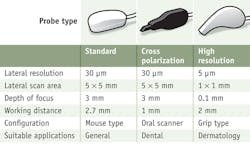
Three different probe formats enable general use (standard mouse type), oral/dental applications (a long neck type), as well as dermatology and oncology (a high-resolution type). (See Fig. 3.) It is the oral/dental probe that incorporates cross-polarization.Software functionality
Software too can have a strong impact on the usability of imaging systems. Features that can enhance this aspect—and promote adoption of the technology—include:
3-D image capture: A 3-D image can be acquired within a couple of seconds and the OCT volume image can be either sliced at the position of interest or exported to a software viewer to provide the clinician with immediate feedback.
Thickness measurement function: This function measures the thickness of layered structures, detects boundaries that appear as peaks in the raw OCT data, and calculates with sub-micron precision distances between multiple peaks.
Surface profiling: A surface profiling function allows the clinician to accurately extract surface or contours of a sample. This function compensates for optical aberrations and scanner non-linearity to generate a distortion-free, 3-D OCT image. A precisely calibrated system can achieve 20 µm accuracy within a 5 mm-sided measurement volume. Combined with thickness measurement, this function enables such new diagnostic possibilities as tumor-growth monitoring.
ACKNOWLEDGEMENTS
The authors thank Dr. Sumi and Dr. Ozawa at the National Center for Geriatrics and Gerontology for collaboration regarding dental applications.
REFERENCES
1. K. Ishibashi et al., J. Dent., doi:10.1016/j.jdent.2011.05.005 (2011).
2. C. Chong et al., IEEE J. Sel. Top. Quant. 14 (1), 235–242 (2008).
Changho Chong is executive director, Daisuke Ogawa is senior scientist, and Jonathan Evans is senior application engineer at Santec Corporation; www.santec.com. Contact Dr. Evans at [email protected].


