PRODUCT FOCUS: OCT systems for ophthalmology

Optical coherence tomography (OCT)—a biomedical imaging technology that can capture three-dimensional (3D) images in-vivo—has emerged as a leading technique in medical diagnosis in the last 21 years since its discovery. And it's no wonder as to why: OCT can produce sub-surface images at resolution >10 μm, which is better than other imaging modalities such as magnetic resonance imaging (MRI) or ultrasound; it is noninvasive; and it does not require any sample preparation or radiation.
To do its job in medical specialties such as ophthalmology, which is the focus here, an OCT system contains a coherent, long-wavelength light source used to scatter off of biological tissue at large angles. Then, an interferometer that typically contains a beamsplitter splits the light source's output into two beams, which then travel and recombine before arriving at a detector. The detector then takes the information collected from the light scattering to produce a 3D image.
So in ophthalmology—which was the first-ever medical specialty to use OCT, by the way—OCT can produce detailed images noninvasively in a way that no other imaging technology can. To learn more about the state of the art and future opportunities for commercial OCT in ophthalmology, I spoke with four developers of OCT systems and components.
Ophthalmic capabilities
"OCT is now indispensible in clinical ophthalmology, and is ubiquitous in research and diagnosis of retinal disease," states Eric Buckland, president and CEO of Bioptigen (Research Triangle Park, NC). In the clinic, it allows clinicians to extract tomographic (or cross-sectional) sections of ocular structures in an "optical biopsy" technique and offers objective measurements of the structures, notes Chris Ritter, Senior Director, OCT Imaging at Carl Zeiss Meditec (Dublin, CA). In diagnosing retinal diseases like glaucoma, retinal thickness and nerve fiber layer thickness measured against normative data are the two most prevalent markers used, adds Buckland. Speaking more to retinal diseases, Jonathan Evans, Ph.D., Director, Optical Systems BU at Santec (Hackensack, NJ), explains that OCT systems also enable before, after, and in-situ monitoring for surgical procedures like capsulotomy and lens fragmentation. These procedures—performed with a precise laser, such as a femtosecond laser—also benefit from OCT as a combination procedure because it allows the surgeon to more precisely place the laser to make its proper cuts.
Anjul Davis, Ph.D., OCT Business Development Manager at Thorlabs (Newton, NJ), notes that OCT's ability to noninvasively examine subsurface structures in the eye enables diagnosis of not only eye disease, but also neurological disease. She also says that the optic nerve is the only nerve in the body that can be accessed through noninvasive imaging. "OCT is also widely used to study diabetes, as research has indicated that diabetes can affect vision," she adds. Diabetic individuals are more likely to develop cataracts at a younger age and twice as likely to develop glaucoma, but diabetic retinopathy (damage to the retina caused by complications from diabetes) is the primary vision problem for diabetics. In this case, OCT systems allow observation of swelling or leakage in the retina.
The need for speed, depth
OCT systems in ophthalmology rely heavily on speed, as faster speed allows for removing motion artifacts—otherwise known as involuntary movements from the patient—and enabling real-time, clearer imaging, says Evans. "OCT in ophthalmology is performed in live patients, largely from the older population, where fixating their eye on one spot is very difficult," explains Davis. Also, faster OCT imaging, she says, enables being able to collect more data in a shorter period of time, such as volumes or blood flow (the Doppler OCT method, in this case).
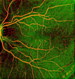
Imaging depth is also important. "OCT is limited in imaging range not just due to absorption and scattering of light through the various layers, but also through fundamental limitations in the total number of points that can be collected in the depth (axial) direction," says Buckland. The ability to image the entire eye (cornea to retina) is desirable, but it requires long-range imaging capability—around >20 mm, explains Davis. So Thorlabs is currently developing a new, high-speed 1050 nm swept source laser specifically for the ophthalmic OCT market. The laser, based on micro-electromechanical systems (MEMS)/vertical-cavity surface-emitting laser (VCSEL) technology, answers the need for speed and depth, with >100 kHz (100,000 lines per second) scanning technology and >25 mm imaging range capability (see Fig. 1). "The availability of such a source for ophthalmic OCT medical device manufacturers means the potential for imaging the entire human eye from cornea to retina," she enthuses.
Novel system achievements
In research, Bioptigen system users study the processes underlying macular degeneration, retinopathy of prematurity, choroidal neovascularization, retinoblastoma, late-gestational stage retinal development, and the use of stem cell therapy in the regeneration of photoreceptors targeted at the cure of retinitis pigmentosa—all using animal models, says Buckland.
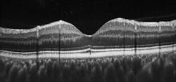
In the clinical space, Bioptigen's Envisu SDOIS C-series OCT system—currently the only OCT system cleared by the FDA for handheld imaging, imaging premature infants, and imaging patients under anesthesia—revealed a focal lesion of the inner segment/outer segment (IS/OS) lines in a patient that was missed using similar systems, notes Buckland. Then, adaptive optics imaging verified that cones were missing in the lesion, which measured 30 μm in diameter. The patient had 20/30 visual acuity, which corresponds to mild vision loss, but he complained of distortions in his vision that couldn't be explained by his physicians. The patient had been in a motor vehicle accident in the past so the OCT system's findings pointed to blunt trauma to the eye, known as commotio retinae (see Fig. 2).What's next?
To make headway in clinical ophthalmology, "point-of-care application [OCT] system cost needs to be reduced," states Evans, and mobility and portability needs to continue to evolve to continue to bring this standard of care to the patient. And as with most imaging-based technologies, OCT resolution and scanning speeds will also continue to evolve upwards, and new wavelengths will likely reveal new aspects and attributes of ocular substructures, says Ritter.
Ritter went on to talk about new clinical applications that will continue to extract more information from OCT 3D data sets to help doctors more efficiently manage disease and provide objective measures for new therapies in development. For example, Carl Zeiss Meditec's Cirrus HD-OCT Advanced Retinal Pigment Epithelium (RPE) Analysis capability enables clinicians to objectively monitor changes associated with dry age-related macular degeneration (AMD), which is the leading cause of blindness in older adults. The system capability can highlight change in RPE elevation area and volume often associated with drusen (extracellular material buildup in the eye that causes lesions), and also identifies and measures the area of transparent regions in the RPE that can develop with geographic atrophy, she explains. "Today, doctors can use this analysis to help them stage dry AMD and track its progression. As new dry AMD drug therapies come to market, this analysis will help doctors track their patient's response to the treatment," she concludes.
About the Author
Lee Dubay
Managing Editor
Lee Dubay is managing editor for Laser Focus World. She is a seasoned editor and content manager with 20 years of experience in B2B media. She specializes in digital/print content management, as well as website analytics, SEO, and social media engagement best practices.

