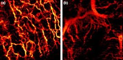
OCT images blood vessels underneath the skin that feed cancer
Researchers from Medical University Vienna (MUW; Vienna, Austria) and Ludwig-Maximilians University (LMU; Munich, Germany) used optical coherence tomography (OCT) to noninvasively map out the network of tiny blood vessels beneath the outer layer of patientsâ skin. Doing so could one day help doctors better diagnose, monitor, and treat skin cancer and other skin conditions.
The researchers tested their system on a range of different skin conditions, including a healthy human palm, allergy-induced eczema on the forearm, dermatitis on the forehead, and two cases of basal cell carcinomaâthe most common type of skin cancerâon the face. Compared to healthy skin, the network of vessels supplying blood to the tested lesions showed significantly altered patterns.
âThe condition of the vascular network carries important information on tissue health and its nutrition,â says Rainer Leitgeb, a researcher at MUW and the studyâs principal investigator. âCurrently, the value of this information is not utilized to its full extent.â
The researchers at MUW are the first to use OCT to visualize the network of blood vessels in human skin that feed cancerous skin lesions. To maximize the quality of the images, the team employed a laser light source developed by the LMU collaborators. The laser enabled unprecedented high-speed imaging and operated at a near-infrared (NIR) wavelength that gave better penetration into skin tissue.
âHigh speed is of paramount importance in order to image lesions in-vivo and in-situ, while minimizing the effect of involuntary patient motion,â explains researcher Cedric Blatter of MUW. The device also shapes the light in a special way forming a Bessel beam, which can reform, or heal, its shape even if portions of it are blocked. The beam enabled the researchers to keep the images in focus across a depth range of approximately 1 mm.
The teamâs images of basal cell carcinoma showed a dense network of unorganized blood vessels, with large vessels abnormally close to the skin surface. The larger vessels branch into secondary vessels that supply blood to energy-hungry tumor regions. The images, together with information about blood flow rates and tissue structure, could yield important insights into the metabolic demands of tumors during different growth stages.
The imaging system shows the most promise for clinical application in the diagnosis and treatment of skin cancer, the researchers believe. âWe hope that improved in-depth diagnosis of tissue alterations due to disease might help to reduce the number of biopsies by providing better guidance,â says Leitgeb. The system could also be used by doctors to assess how quickly a tumor is likely to grow and spread, as well as to monitor the effectiveness of treatments such as topical chemotherapy. âTreatment monitoring may also be expanded toward inflammatory and auto-immune related dermatological conditions,â Blatter notes.
Going forward, the researchers would like to increase the field of view of the device so that they can image the full lesion along with its border to healthy tissue. They are also working on speeding up the post-processing of the optical signal to enable live vasculature display and improving the portability of the system, which currently occupies an area about half the size of an office desk. âWe believe that in the future our method will help to simplify non-invasive dermatological in-vivo diagnostics and allow for in-depth treatment monitoring,â says Blatter.
The work has been published in Biomedical Optics Express; for more information, please visit http://www.opticsinfobase.org/boe/abstract.cfm?uri=boe-3-10-2636.
-----
Follow us on Twitter, 'like' us on Facebook, and join our group on LinkedIn
Laser Focus World has gone mobile: Get all of the mobile-friendly options here.
Subscribe now to BioOptics World magazine; it's free!
