Endoscopic OCT system could have use in minimally invasive joint surgery
A team of researchers at Duke University's Pratt School of Engineering (Durham, NC) has developed a way to use optical coherence tomography (OCT) imaging in areas of the body that are difficult to reach, such as joints. The work is promising for bringing OCT imaging to new surgical and medical applications.
OCT can image structures measured in microns, allowing clinicians to see subtle changes in tissue that might indicate disease or damage. Although OCT is now the standard of care in ophthalmology, making a high-quality OCT instrument compact enough for use inside the body has been challenging.
The researchers used a rigid, 4 mm borescope (essentially a thin tube of lenses) to deliver the infrared light necessary to perform OCT. The borescope makes the beam delivery portion of the device very slim without sacrificing imaging performance.
"We saw a need for OCT image guidance in arthroscopic surgery, a minimally invasive procedure that uses an endoscope to address joint damage," says Evan T. Jelly, research team leader. "We took the low-cost OCT imaging platform we previously developed and adapted it to meet the requirements of this application."
Working with Adam Wax, Professor of Biomedical Engineering at Duke University, Jelly and his research team previously developed OCT systems that are far lower in cost than traditional systems. To make an OCT system that could be used to assess the health of cartilage in a joint, they created an endoscopic delivery system that uses a prototype rigid borescope to relay the image from the tissue to a fiber-optic connection. The instrument's narrow front viewing section allows it to reach structures and cavities that aren't accessible with the larger portion of the device that scans the patient.
Related: Ultralight, low-cost OCT scanner screens for retinal diseases
The researchers designed the endoscopic OCT instrument to provide real-time quantitative information on cartilage thickness without requiring a clinician to cut or damage the tissue. This type of analysis is important for painful joint conditions such as osteoarthritis, which develop when cartilage wears down and becomes thinner.
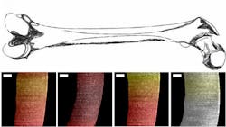
The researchers tested their OCT instrument by using it to measure the thickness of cartilage in pig knees. Because pig cartilage is similar to that of humans, this provided a preliminary idea of how the device would perform in humans. The system was able to accurately identify the bone-cartilage interface for samples that were less than 1.1 mm thick.
With further development, the device could one day allow clinicians to offer less invasive treatment of joint problems. However, the researchers still need to demonstrate that it can image thicker samples since human cartilage is slightly thicker than pig cartilage. They also want to further improve the ergonomics for use during surgery.
"By developing a new portable, low-cost version of OCT, we show that the success of this imaging approach will no longer be limited to ophthalmology applications," says Jelly. "With some engineering expertise, this OCT platform can be adapted to fit a wide range of clinical needs."
Full details of the work appear in the journal Optics Letters.
Source: Duke University Pratt School of Engineering press release
Got biophotonics-related news to share with us? Contact Lee Dubay, Associate Editor, BioOptics World
Get even more news like this delivered right to your inbox
