30-minute blood test uses microarray to detect E. coli bacteria
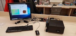
With a goal to reduce the mortality rate from sepsis by more than 70%, a team of European scientists has developed a microarray detector that uses a tiny blood sample to produce results in less than 30 minutes. Current techniques for detecting sepsis can take hours or even days to produce the results and diagnosis.
Programmed to detect proteins and E. coli, one of the deadly bacteria that can cause the human body to go into septic shock, the detector then uses light to look for specific biomarkers (the telltale signs or an indicator of a disease) that are as small as few nanometers in size.
The rapid microarray detector looks at a small blood sample taken from a thumb or forefinger. The patient's blood sample is then separated in a centrifuge so that a clinician can examine the plasma, the part of the blood sample where all the proteins are contained.
"The optical readout of the sample can be completed in one minute, allowing us to deliver results in 30 minutes from start to finish," says project coordinator Roland Terborg. "This is much faster than methods currently in use. With a condition like sepsis where time is crucial, this device looks set to prevent thousands of deaths every year that could easily have been avoided."
Developed by the "Scalable point-of-care and label-free microarray platform for rapid detection of Sepsis" (RAIS) project, the project was coordinated by The Institute of Photonic Sciences (ICFO; Barcelona, Spain) and is a success story for the Photonics Public Private Partnership.
Related: Microscope detects one million-plus biomarkers for sepsis in 30 minutes
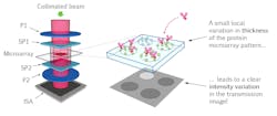
The sepsis detector uses photonics to make a clear and accurate diagnosis. The plasma sample flows over a microarray, a collection of tiny spots containing specific antibodies on a nanostructured gold slide. Two light beams are then shone through the full microarray, with one of them passing through the sample, while the other one goes through the clear part of the slide, acting as a reference. The beams passing through the biomarker and the clear regions on the slide are then checked for any changes in intensity."Depending on the amount and type of biomarker attached to each antibody, we obtain a unique image: a signature pattern if you like," Terborg explains. "The image patterns tell us what is present in the plasma sample, which we then record with a CMOS sensor—the same technology used in a digital camera that converts light into electrons."
The rapid detection of sepsis could save the healthcare system several tens of billion euros per year due to the decrease of hospital stays, and the reduction of unnecessary drug usage and associated insurance costs. The device could also be extended to perform on other types of disease screening or multiple simultaneous diagnoses, especially those requiring a rapid detection of large numbers of biochemical targets (more than 1 million) on a single microarray.
Preclinical trials have already begun at Vall d'Hebron University Hospital (also in Barcelona), where the device has been in operation since 2018. Clinical trials are expected to take place at the end of 2019.
For more information, please visit photonics21.org.

