Optical probes overcome light-scattering issue in deep-brain imaging
Light scattering and absorption in neural tissue cause light penetration to be extremely short, making it impossible to employ free-space optical methods like optogenetics to probe brain regions deeper than about 2 mm.
Related: Ten years after - Optogenetics progresses in clinical trials
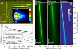
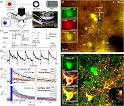
Recognizing this, a team of researchers from the Calfornia Institute of Technology (Caltech; Pasadena, CA), the Baylor College of Medicine (Waco, TX), and Stanford University (CA) has developed a new approach that combines nanophotonics and microelectromechanical systems (MEMS) in an implantable, ultra-narrow, silicon-based photonic probe to deliver light deep within brain tissues. This minimally invasive technique avoids major tissue displacement during implantation.
Using optogenetic techniques, a protein in the brain serves as a sensory photoreceptor and can be controlled by specific wavelengths of light. These combined techniques provide a new approach to stimulating brain circuits with remarkable resolution, enabling observation and control of individual neurons.
These breakthroughs present widespread and promising applications for the neuroscience and neuromedical research communities. From characterizing the role of specific neurons and identifying neural circuits responsible for behavior to enabling new methods of operant conditioning through reward-induced circuit activations, optogenetics has become a new path for neuroscientists seeking advances in research capabilities.
Full details of the work appear in the journal Neurophotonics (open access); for more information, please visit http://dx.doi.org/10.1117/1.nph.4.1.011002.
BioOptics World Editors
We edited the content of this article, which was contributed by outside sources, to fit our style and substance requirements. (Editor’s Note: BioOptics World has folded as a brand and is now part of Laser Focus World, effective in 2022.)

