Raman spectroscopy following laser ablation shows promise for cancer surgery
In a study, researchers at Florida Atlantic University (FAU; Boca Raton, FL) used Raman spectroscopy to distinguish normal from skin tissue with cancer following high-powered laser ablation.
Related: Laser ablation helps to home in on cancer cells
This is reportedly the first time that Raman spectroscopy has been successfully used to detect cancerous tissue following laser ablation, setting the stage to use this method as a guide for laser surgery. Scientists hope to one day employ Raman spectroscopy clinically in tandem with laser-ablative removal of skin cancer and possibly even other forms of cancers as well.
Cancerous tissue is diagnosed using a Raman laser and the cancerous tissue is removed in a very precise, localized fashion using an ablation laser. This type of Raman-based technique for skin cancer diagnosis and treatment, although not yet fully developed, may one day obviate the need for a more costly and time-consuming treatment method like Mohs micrographic surgery, at least in some, if not all, clinical cases.
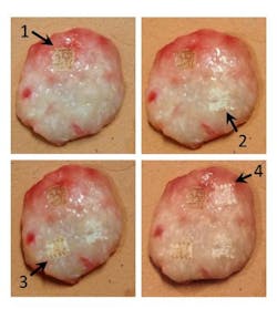
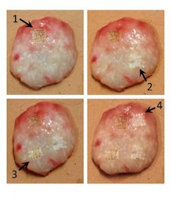
This study combined laser ablation for highly precise, hemostatic removal of cancerous tissue, with Raman spectroscopy to objectively and non-destructively probe the ablated tissue area in situ for any remaining cancer to remove. Raman spectra were collected from partially ablated normal and squamous cell carcinoma samples, and a spectral classification model based on principal component analysis with logistic regression correctly identified spectra from residual cancerous tissue with 95-percent sensitivity and 100-percent specificity.
"Successful clinical implementation of the proposed surgical method could greatly enhance the speed and effectiveness of skin cancer treatment, especially if real-time analysis of the process were developed," says Andrew C. Terentis, Ph.D., lead scientist of the study and an associate professor of chemistry and biochemistry in FAU's Charles E. Schmidt College of Science.
Mohs micrographic surgery is currently the gold standard technique for skin cancer removal because it provides high cure rates while removing the least amount of healthy tissue. However, it is time consuming, resource-intensive, and the intraoperative evaluation of the excised tissue sections by histopathology is subjective. With Mohs surgery, the physician serves as surgeon, pathologist, and reconstructive surgeon and relies on the accuracy of a microscope to trace and ensure removal of skin cancer down to its roots. This procedure allows dermatologists, trained in Mohs Surgery, to see beyond the visible disease, and to precisely identify and remove the entire tumor, leaving healthy tissue unharmed.
Terentis' collaborators include co-author John Strasswimmer, MD, Ph.D., affiliate faculty in FAU's Charles E. Schmidt College of Medicine, and a skin cancer specialist and director of the Melanoma and Cutaneous Oncology Program at the Lynn Cancer Institute and Moffitt Cancer Network. Partial funding was provided by the Sinai Hospital (Detroit, MI).
"When a surgeon removes a cancer, whether it be with Mohs surgery for skin cancer or a surgeon using a robot in a modern operating room for abdominal cancer, the surgeon must rely on vision and touch to help decide initially how much tissue to remove," says Strawswimmer. "This new work with Professor Terentis sets the stage for us to have an automatic laser to vaporize cancer and the Raman spectroscopy to tell us when to stop the vaporization process. This is particularly important in areas that we can access with the laser beam such as the lungs or inside the liver that are otherwise very difficult to access with traditional surgery. We designed this study with skin cancer because it is a very straightforward model study and the number of skin cancer patients is increasing at an exponential rate."
Full details of the work appear in the journal Lasers in Surgery and Medicine; for more information, please visit http://dx.doi.org/10.1002/lsm.22288.
-----
Follow us on Twitter, 'like' us on Facebook, connect with us on Google+, and join our group on LinkedIn
Subscribe now to BioOptics World magazine; it's free!

