Near-infrared spectroscopy improves osteoarthritis evaluation
A researcher at the University of Eastern Finland (Joensuu, Kuopio, Finland) has developed an arthroscopic near-infrared spectroscopy (NIRS) probe for evaluating articular cartilage and subchondral bone structure and composition. The probe enables enhanced detection of cartilage injuries due to osteoarthritis, as well as evaluation of the integrity of the surrounding tissue. The availability of comprehensive information on the health of joint tissues could substantially enhance the treatment outcome of arthroscopic intervention.
Clinical application of NIRS is now possible, thanks to better availability of computational power along with state-of-the-art mathematical modeling methods such as neural networks. With these methods, the relationship between the absorption of NIR light and tissue properties can be determined. This enables reliable determination of articular cartilage stiffness and subchondral bone mineral density—changes in these tissue properties are prognostic indicators of osteoarthritis.
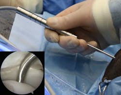
The novel arthroscopic probe in an equine knee joint in vivo with the probe tip in contact with cartilage surface (inset). (Photo credit: Jaakko Sarin)
Since NIRS is not optimal for imaging of tissues, arthroscopically applicable imaging techniques such as optical coherence tomography (OCT) and ultrasound imaging were also used in the study. These techniques have been previously applied in intravascular imaging via specialized 1-mm-diameter catheters, which are therefore well suited for imaging narrow joint cavities. The study compared the reliability of these techniques for evaluation of chondral injuries with that of conventional arthroscopic evaluation.
"Optical coherence tomography was superior to conventional arthroscopy and ultrasound imaging. In contrast to conventional arthroscopic evaluation, optical coherence tomography and ultrasound imaging provide information on inner structures of cartilage and enable, for example, detection of cartilage detachment from subchondral bone," explains Jaakko Sarin, the researcher from the University of Eastern Finland who led the development.
Full details of the work appear in the journal Scientific Reports.
