Apps are changing the world of microscopy. So if "Angry Birds" is what comes to mind when you hear the word "app," you need to update your downloads!
Already fueling new approaches in education at institutions such as Yale University (New Haven, CT), easy-to-use apps for tablets and smartphones are getting set to propel research as well. Let's look at how releases from vendors such as Leica Microsystems (Wetzlar, Germany), Olympus Scientific Solutions Americas (Waltham, MA), and Carl Zeiss Microscopy (Thornwood, NY) are changing processes in microscopy and promising to provide specialized capabilities in the future.
Speeding setup
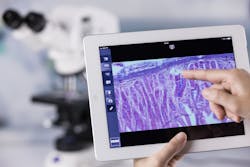
"In a course that introduces students to the model organisms used in biological research, we use microscopes quite a bit," says Maria Moreno, a research scientist in the biology department at Yale. To share images around the class, Moreno uses Leica's DMshare—a free app that works with particular Leica microscopes and cameras, plus a data-transfer system. With this app, an image on a microscope can be captured with a Leica camera and then viewed wirelessly on an iPad (see Fig. 1). "We can take pictures with this, and annotate them," says Moreno.
The current version of DMshare allows eight iPads to be connected to one scope. "But you can create an external network and have any number of users," Moreno says. "I can have all of my students visit a router connected to the microscope, and they can see what I'm showing."
Beyond making it easier to teach a lab class that uses microscopes, DMshare changes the experience. "It has revolutionized how things go in my classroom," Moreno says. "I can show my students exactly how to set up the microscope with the iPad, and they can see what it looks like when the setup is right and wrong." She says that helping everyone get his or her microscopes set up right used to take a lot of her teaching time. "Now," she says, "it takes no more than 15 minutes because everyone can see the same thing."
Moreno says the app is "a really great tool" and yearns for just one improvement: "I wish that my students could use this app to take movies," she says. That advance could lie just ahead. According to Karin Schwab, product manager for DMshare at Leica Microsystems, "We frequently update this app. We've just recently implemented more cameras, imaging systems like the Leica DMS3000, and even added a fluorescence camera."
Beyond education, says Schwab, DMshare benefits research because it makes it easy for scientists to share and discuss results—even when they are away from the microscope.
Cutting the curve
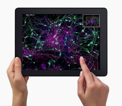
Increasing accessibility seems to be a major benefit of apps from microscope makers, in fact. Kristen Orlowski, product marketing manager, light microscopes at Carl Zeiss Microscopy, says, "Our Labscope app works with two cameras, including one that's in the binocular head of an educational microscope." This app, which works with an iPad or iPhone, "takes the learning curve out of digital imaging," Orlowski explains (see Fig. 2). "You don't need any fancy software that often intimidates people." She adds, "Everybody is familiar with apps."Much of the simplicity comes from the app environment, where things just seem to make sense. With Labscope, for instance, a student just hits a button that looks like a camera, and it takes a picture.
This app is also free, so it can be used in almost any classroom that has the right microscope or camera. "In a classroom of even 15 or 16 microscopes," says Orlowski, "the instructor can tap into any student's scope and see exactly what that student is looking at." Then, she says, "If you see something very interesting, the instructor can hit a button to put that scope's image on a shared screen, instead of lining everyone up at the scope."
Centered on sharing
"With really high-resolution images," says Christopher Higgins, VS120 applications manager for whole-slide imaging at Olympus Scientific Solutions Americas, "you can't just push them out in e-mails to share." To share images around the world, Olympus developed its OlyVIA app. "We launched it at first for education," Higgins says, "but we can also support confocal images, time lapse, multi-z focus, and more." So this app also offers many research possibilities.
For Olympus, volume started the process. As Higgins explains, "We came up with the idea for this application to share these amazing multi-billion-pixel images that the VS120 and our other systems can generate." He adds, "The Olympus OlyVIA mobile iPad app allows users to gain access to and make use of their entire library of virtual microscope images from anywhere in the world. With a click, a tap, or a finger pinch, they can select, pan, zoom, and work with their images at various magnifications, taking full advantage of the portability and the user-friendliness of their iPads." Like the other apps mentioned here, this one is free (see Fig. 3).This app works with a range of Olympus technology. Higgins says, "The OlyVIA app is designed for use with images captured with Olympus imaging systems and shared with others using Olympus Net Image Server v.2.7 software."
This app can be used in teaching or research. In education, says Higgins, "Teaching and training are streamlined thanks to the easy upload of stored images, which are provided to students for study and evaluation." He also points out that it's useful in research, where "huge image files are shared and consulted upon by colleagues working remotely."
Carl Zeiss offers a similar app called Zen Browser. As Orlowski says, it "allows users to access any Zeiss system image on their server from a mobile device." She adds, "The software allows for an unlimited number of users to have access to the images stored."
Specialization coming up
In the future, says Orlowski, "You will start seeing apps tailored to very specific things, like finding specific structures or looking for particular features of a cell." Plus, she points out that apps are very easy to update. That, too, should contribute to even more intriguing microscope apps ahead.
Mike May | Contributing Editor, BioOptics World
Mike May writes about instrumentation design and application for BioOptics World. He earned his Ph.D. in neurobiology and behavior from Cornell University and is a member of Sigma Xi: The Scientific Research Society. He has written two books and scores of articles in the field of biomedicine.


