PHOTOACOUSTICS/BREAST CANCER: Photoacoustic mammoscopy aims for safer, earlier breast cancer screening

Nov. 18, 2013
The photoacoustic mammoscope represents an entirely new way of imaging the breast, and hopes to detect cancer earlier than current methods.

The photoacoustic mammoscope represents an entirely new way of imaging the breast, and hopes to detect cancer earlier than current methods. Instead of potentially hazardous x-rays, which are used in traditional mammography, the photoacoustic mammoscope, designed by researchers in the University of Twente (The Netherlands) Biomedical Photonic Imaging group, uses a combination of benign infrared (IR) light and ultrasound to create a 3D map.

The technique delivers to tissue IR light in billionth-of-a-second pulses. Relatively high absorption by blood increases the temperature of blood vessels, which expand slightly but quickly. While imperceptible to the patient, this expansion generates detectable ultrasound waves that enable formation of a 3D map of breast vasculature. Since cancer tumors have more blood vessels than the surrounding tissue, they are distinguishable in this image.

A small clinical trial showed in 2012 that an earlier version of the technology could successfully image breast cancer in women. In future versions, the researchers expect to improve resolution and add the capability to image using several different wavelengths simultaneously, which promises to improve its ability to detect cancer.

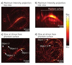
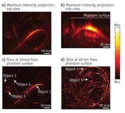
Top (a) and side (b) views show a 3D-reconstructed tissue phantom, the background of which mimics normal breast tissue, while several embedded objects mimic blood vessels and tumors. In two image slices (c and d) taken with the new device, locations of five objects are indicated with arrows: objects 1 and 2 are blood vessel mimics, while objects 3, 4, and 5 are phantom tumors. (Image courtesy of Wenfeng Xia, Biomedical Photonic Imaging group, University of Twente)

A commercial instrument using this technology would likely cost much less than MRI and x-ray instruments, and would be more in line with ultrasound systems, said Wenfeng Xia, first author on a paper describing the system.1 The next step is to prepare for larger clinical trials; the final prototype will be ready for testing in 2014.

1. W. Xia et al., Biomed. Opt. Exp., 4, 11, 2555–2569 (2013).
Sponsored Recommendations

Demonstrating Flexible, Powerful 5-axis Laser Micromachining

Sept. 18, 2024
Five-axis scan heads offer fast and flexible solutions for generating precise holes, contoured slots and other geometries with fully defined cross sections. With a suitable system...

Enhance Your Experiments with Chroma's Spectra Viewer

Sept. 5, 2024
Visualize and compare fluorescence spectra with our interactive Spectra Viewer tool. Easily compare and optimize filters and fluorochromes for your experiments with this intuitive...

Optical Filter Orientation Guide

Sept. 5, 2024
Ensure optimal performance of your optical filters with our Orientation Guide. Learn the correct placement and handling techniques to maximize light transmission and filter efficiency...

Ensure Optimal Performance with Shortpass Filters

Sept. 5, 2024
Achieve precise wavelength blocking with our Shortpass Filters. Ideal for applications requiring effective light transmission and cutoff, these filters ensure optimal performance...

Voice your opinion!

To join the conversation, and become an exclusive member of Laser Focus World, create an account today!