Optical imaging technique helps diagnose, monitor major diabetes complication
Columbia University (New York, NY) researchers have developed a noninvasive optical imaging device that could ease diagnosis and monitoring of peripheral arterial disease (PAD), which is a serious complication of diabetes. PAD is marked by a narrowing of the arteries caused by plaque accumulation, frequently resulting in insufficient blood flow to the bodyâs extremities and increasing a personâs risk for heart attack and stroke.
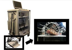
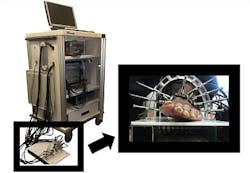
The imaging device employs dynamic diffuse optical tomography (DDOT) imaging, which uses near-infrared (NIR) light to map the concentration of hemoglobin in the bodyâs tissue. The technique can reveal how effectively blood is flowing to patientsâ hands and feet.
Right now, there are no good methods in place to assess and monitor PAD in diabetic patients, explains Andreas Hielscher, Ph.D., professor of Biomedical and Electrical Engineering and Radiology, and director of the Biophotonics and Optical Radiology Laboratory at Columbia University.
âPatients with PAD experience foot pain, called âclaudication,â while walking,â explains Gautam Shrikhande, MD, assistant professor of surgery and director of the Vascular Laboratory at Columbiaâs Medical Center. âThis pain continues, even at rest, as the disease progresses. In more advanced stages, PAD patients develop sores or ulcers that wonât heal. Then, cell death, a.k.a. âgangrene,â occurs and amputation is often the only solution. Itâs extremely important to diagnose PAD early, because medication and lifestyle changes can alleviate the disease.â
The researchers used DDOT to detect PAD in the lower extremities, according to Michael Khalil, a Ph.D. candidate working with Hielscher at Columbia. DDOT, unlike other methods, can provide maps of oxy, deoxy, and total hemoglobin (the protein that carries oxygen in the blood) concentration throughout the foot and identify problematic regions that require intervention, he says.
âUsing instrumentation for fast image acquisition lets us observe blood volume over time in response to stimulus such as a pressure cuff occlusion or blockage,â said Hielscher.
To map and monitor the presence of hemoglobin, the DDOT device shines red and NIR light at different angles around areas that are susceptible to arterial disease. Then, these specific wavelengths of light are then absorbed or scattered, depending on the concentration of hemoglobin.
âIn the case of tissue, light is absorbed by hemoglobin. Since hemoglobin is the main protein in blood, we can image blood concentrations within the foot without using a contrast agent,â Hielscher points out. Contrast agents pose the risk of renal failure in some cases, so the ability to monitor PAD without using a contrast agent is a great advantage.
Khalil, Hielscher, and colleagues hope to commercialize the device and bring it into clinics within the next 3 years.
The work has been published in Biomedical Optics Express; for more information, please visit http://www.opticsinfobase.org/boe/abstract.cfm?uri=boe-3-9-2288.
-----
Follow us on Twitter, 'like' us on Facebook, and join our group on LinkedIn
Laser Focus World has gone mobile: Get all of the mobile-friendly options here.
Subscribe now to BioOptics World magazine; it's free!


