OPTOACOUSTICS/MEDICAL IMAGING STANDARDS: Getting quantitative data from photoacoustic images
Photoacoustic imaging is an emerging biomedical imaging modality capable of visualizing optically absorbing structures with high resolution (sub-50 microns) to centimeter depths in optically scattering soft tissue. The visualization of blood vessels and capillaries has been a priority for researchers for two reasons. First, because the hemoglobin naturally present in red blood cells acts as a source of optical contrast so no external contrast agent is required. Also, because the absorption spectrum of hemoglobin depends on whether it is carrying oxygen (oxy- or deoxy-hemoglobin), it should be possible–by making measurements at multiple wavelengths–to obtain a 3D map showing the vessels and their level of blood oxygenation.
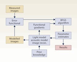
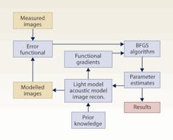
Images simulated with a computational model are matched to the measured images; and model input parameters are adjusted until they agree. The method makes use of the Broyden-Fletcher-Goldfarb-Shanno (BFGS) algorithm. (Diagram courtesy of Ben Cox)
However, this task turns out not to be as straightforward as it sounds. Although absorption provides contrast, a photoacoustic image is not proportional to the absorption coefficient but is a product of the absorption and the light fluence distribution. As the light distribution will be different at each wavelength, a ratio of photoacoustic images obtained at different wavelengths does not correspond to a ratio of absorption, so in most circumstances cannot be used for the direct calculation of blood oxygenation. Furthermore, calculating the light distribution and removing it from the image is not a simple task as it depends on the very absorbers one is trying to estimate.
In a paper published by the Optical Society (OSA), researchers in the Photoacoustic Imaging Group at University College London (UCL, England), led by Professor Paul Beard, describe progress they have made to overcome this problem.1 The group showed that the concentrations of two chromophores (analogous to oxy- and deoxy-hemoglobin) and their ratio (analogous to blood oxygenation) can be estimated from experimentally measured photoacoustic images by using an iterative model-based nonlinear inversion. This works by matching images simulated with a computational model to the measured images, and adjusting the model input parameters (the chromophore concentrations), until they agree.
The implications of this result go beyond the measurement of blood oxygenation. The ability to determine the absolute concentrations of absorbers from photoacoustic images is a significant step towards high resolution, quantitative, imaging of molecularly targeted or genetically expressed contrast agents amidst a background of endogenous tissue chromophores. Such an imaging tool would find widespread use in biomedicine and life sciences research.
–Ben Cox
Ben Cox is Research Fellow, Dept. Medical Physics and Bioengineering, UCL, London UK
Reference
- J. Laufer et al., Applied Optics 49 (8), 1219-1233 (2010).
More Brand Name Current Issue Articles
More Brand Name Archives Issue Articles

