On opening day of the Biomedical Optics Symposium (BiOS) at SPIE Photonics West 2020, an industry session showcasing commercial developments in optical coherence tomography (OCT) from five developers heralded dramatic changes (like widespread application facilitated by small, low-cost systems), and clinical impacts promised by cumulative technological developments.1
Scan speed, FOV, and system size
Carl Zeiss Meditec (Jena, Germany) has been at the forefront of OCT technology from the beginning when the ophthalmic instrumentation maker brought the first time-domain OCT system to market and then, over almost 20 years, played a key role in making OCT the standard of care in ophthalmology.
Tilman Schmoll, principal investigator of the Zeiss Lab at the Medical University of Vienna (Austria), described OCT’s progress, saying the newest OCT instruments operate roughly 2000X faster than first-generation systems—and added that ZEISS expects this exponential growth to continue. (By the way, speed increases did not come at the expense of image quality, which has also improved.)
So far, Schmoll said, speed increases have mainly enabled wider fields of view, so that total acquisition time has remained roughly constant. But, the technology is now capable of speeds that allow extensions such as OCT angiography (OCTA), in which multiple scans, acquired at the same location, enable detection of changes such as blood cells moving through retinal capillaries.
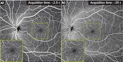
Most current commercial OCT systems offer scan patterns from 3 × 3 mm to 12 × 12 mm field of view (FOV) with 3 mm scan depth and lateral resolution of 20 to 25 µm. Acquisition speed is the main factor limiting FOV width—in fact, wide-field OCTA is usually achieved by combining scans (for example, two partially overlapping 15 × 9 scans are registered and blended to produce a 15 × 15 montage. But, new technology now allows a single 15 × 15 scan at 200 kHz, which can replace the co-registered 15 × 9 images scanned at 100 kHz while maintaining image resolution. In addition, ZEISS researchers say the new scan modality can reduce acquisition time by another ~25% because the mitigated risk of eye motion decreases the need for rescanning.
An add-on lens enables single-shot capture of a 24 × 24 OCTA scan—with 27 µm resolution and a FOV of 90°—equivalent to the output of ZEISS’s CLARUS 500 wide field fundus camera, a development that promises to facilitate not only monitoring and treatment in patients with diabetes, but also understanding of the disease.
ZEISS’s R&D team has proven additional clinical value for widefield imaging at megahertz speeds, as described in a conference presentation (see Fig. 1).2
Schmoll discussed ZEISS’s role in the Ophthalmic OCT on a Chip project, a five-year project due to conclude at the end of 2020.3 The consortium aims to apply photonic integrated circuits (PICs) to drive down OCT system size and cost, and through integration with CMOS electronics, it promises mass fabrication for maintenance- and alignment-free devices that will enable new applications in point-of-care diagnostics and digital medicine. A conference paper presented the project’s first in vivo results with clinically useful image quality, and described two systems based on the technology.4
Zeiss is also participating in another five-year project, which launched in January 2020. Called Handheld OCT, it aims to facilitate a new generation of 1060 nm devices with dramatic increase in imaging speed, and decreases in size and cost compared to the current state-of-the-art.5 As described in another conference paper, the consortium aims to achieve these goals through “monolithic integration of silicon nitride optical waveguides, germanium photodiodes, and micro-optics combined with the hybrid integration of a novel compact all-semiconductor akinetic swept source,” and has an ultimate objective to promote widespread adoption of OCT in point-of-care diagnostics and diagnostic-driven therapy of retinal pathologies.6
Smaller, faster, more affordable, more capable
Another company that has been a powerhouse in OCT is Thorlabs (Newton, NJ), a developer and manufacturer of a broad portfolio of OCT products, from components (including a range of light sources, optical and mechanical items, and detectors) to modular subsystems and full-on turnkey systems for more than 15 years. The company has manufacturing facilities for optomechanics, optics and optical coatings, optoelectronics, optical fibers, and semiconductor wafer growth and fabrication, and works collaboratively with researchers, industry partners, and customers to advance OCT.
Thorlabs’ component business serves not only its own supply line, but also the OEM market—therefore, it offers a range of OEM products as well as engineering for integration of customized OCT modules into complex host systems.
In summarizing recent work at Thorlabs, lead VCSEL applications scientist Shahid Islam noted miniaturization and cost reduction of key components. He previewed a number of new products, including a multimodal version of the Telesto OCT system (for release in 2020) that provides simultaneous acquisition of OCT and fluorescence imagery. Also new are wideband fiber-optic couplers designed for splitting and mixing optical signals with specific coupling ratios, which feature a broad spectral range and minimal spectral dependency, for easy integration into OCT systems, and OCT balanced detectors with fast monitor output covering various wavelengths and bandwidths. And, new additions to Thorlabs’ indium phosphide (InP)- and gallium arsenide (GaAs)-based Booster Optical Amplifier (BOA) line mean that offerings now span 980 to 1650 nm.
And while Thorlabs offers a range of light sources for OCT, its flagship illumination product is its tunable MEMS-VCSEL swept wavelength laser source, featuring >100 mm coherence length and active power control to maintain constant lifetime output. (By the way, MEMS stands for microelectromechanical system and VCSEL is vertical-cavity surface-emitting laser.) VCSEL-based OCT provides a combination of high speed, operational flexibility, and long range that promises new applications in biomedicine and beyond. The technology, now offered in both 1060 and 1300 nm wavelengths, now offers fast, switchable, multiple sweep modes, wherein a laser can have up to three completely independent sweep modes and up to three (optional) Mach-Zehnder interferometer (MZI) k-clocks to enable multi-rate, multi-MZI operation.
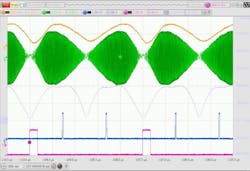
A new capability of the MEMS-VCSEL source is optimized bidirectional sweep, enabling up to 1.2 MHz effective sweep rate (2–600 kHz unidirectionally; see Fig. 2). Like Zeiss, Thorlabs is working on even greater speeds, and in the technology’s evolution from Gen1 to Gen2, the sweep rate is expected to reach at least 1 MHz, or >2 MHz bidirectionally. In addition, Gen 2 promises a modular design to accommodate a broad range of customization.The company recently announced benchtop/turnkey MEMS-VCSEL swept sources (including 1060 and 1300 nm variants with a range of sweep rates for different imaging depth ranges) offering dramatically improved imaging depth with almost no loss of OCT sensitivity.
Thorlabs’ VCSEL technology was highlighted in three conference presentations, including two focused on the 1050 nm technology (one was an invited talk7 and the other was part of the Novel Light Sources session8), while another invited talk covered continuous-wave mid-infrared VCSELs.9
Vastly more affordable, right now
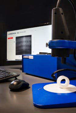
Duke University professor Adam Wax founded Lumedica (at which he serves as Chief Science Officer) expressly for the purpose of making OCT vastly more affordable. He was inspired by the fact that current standard OCT systems cost ~$35,000, a price point that limits the technology’s reach into new clinical areas and under-resourced locations. In the BiOS exhibit hall, Lumedica displayed its full product line of low-cost OCT systems designed to support researchers, educators, clinicians, and OEMs. Among the offerings is the OQ Labscope 2.0, which provides a complete system with engine and scanner for just $9995 and offers such options as increased speed (up to 80 kHz), resolution (to 2 mm axial) and extended depth penetration (1310 nm; see Fig. 3). The company’s second-generation offering is a clinical OCT system that is even more affordable and portable (weighing just 4 lbs., including integrated PC, touchscreen, and handheld probe).Clinical applications for the low-cost OCT technology include ophthalmology, of course, and an integrated Lumedica system was investigated in a recent clinical study10 that showed the system approaches the performance of standard commercial ophthalmic systems. Specifically, it provides 94.4% of the contrast-to-noise ratio of Heidelberg Engineering’s Spectralis instrument—at 10X less cost and 10X smaller size.
Adaptations for other applications, with cost and size to meet point-of-care needs, are reported in other journal papers:
- Dermatology, with dual-axis OCT. Surgical models show the ability to see diagnostic features through skin11
- Gastroenterology, specifically endoscopic imaging of Barrett’s esophagus epithelium (a current clinical study uses a 3D-printed paddle probe)
- Gynecology, for imaging of the so-called transformation (T)-zone of the cervix, the area most likely to develop abnormal cells (a clinical study is underway)12
- Orthopedic measurement of articular cartilage thickness, with potential for integration into live imaging to guide surgery (no standard intraoperative method exists)13
- Neurodegeneration, including multimodal screening and search for biomarkers of Alzheimer’s-related retinal change. The approach leverages another one of the Duke researchers’ areas of focus, angle-resolved low-coherence interferometry (a/LCI), along with fluorescence and histology in combination with OCT.14
- Arthroscopy and general surgery: novel applications using a system designed for OCT imaging through a commercially available rigid borescope.
Application: Ear infections
PhotoniCare cofounder and CEO Ryan Shelton described an OCT-based handheld diagnostic platform for front-line healthcare with an initial application of providing the first objective, quantitative, and noninvasive assessment of middle ear infections, the leading cause of hearing loss, surgeries, and antibiotic use in U.S. children. Shelton titled his talk “Through the Curtain” because, he explained, standard technologies used to assess ear infections look at the “curtain” (that is, the tympanic membrane, a.k.a. eardrum or TM), offering no way to actually visualize infection behind it. What’s more, current techniques are subjective, resulting in a 50% misdiagnosis rate for primary care providers.
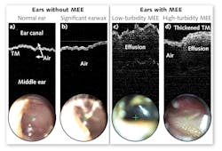
By contrast, PhotoniCare’s handheld TOMi Scope provides unprecedented views of the middle ear, enabling quantification of effusion and purulence, and visualization of the TM’s structure and thickness, which enables disease classification (see Fig. 4). A recent study done in the lab of Stephen Boppart at the University of Illinois at Urbana-Champaign15 reports 98% accuracy resulting from the use of machine learning for automated classification, for which the company earned an NIH grant—though a 70-patient clinical study measuring fluid detection capability (for which the system has earned U.S. FDA 510(k) clearance) showed more than 90% sensitivity, specificity, and accuracy, and high intrareader agreement.16 PhotoniCare expects expanded 510(k) clearance for advanced algorithms to facilitate decision support.PhotoniCare’s model estimates potential cost avoidance at $150–250 per use (amounting to a substantial figure when multiplied by the large number of cases). The company currently has several letters of intent from customers (with others pending), and has obtained two CPT codes for insurance billing.
Application: Cardiovascular disease
The world’s leading cause of death, according to the World Health Organization, is cardiovascular disease (CVD), wherein cholesterol in the bloodstream, deposited onto vessel walls, creates lipid pools. Covered by very thin tissue, a lipid pool can rupture, triggering thrombosis and restricting blood flow, which damages the heart. A common treatment is stenting, but many complications can result from this procedure, and while image guidance has been shown to improve stenting outcomes, it is used in only 15% of cases worldwide.
The current standard of care consists of pharmacologic treatment and percutaneous coronary intervention (PCI). PCI begins with incision in the femoral artery and placement of a catheter that is guided by angiography to the region of interest, where a stent is placed and expanded using a balloon. Technology developed by SpectraWAVE, a startup based on the work of Gary Tearney’s lab at Harvard Medical School and Massachusetts General Hospital (Boston, MA), aims to assist with guidance of PCI—and also to improve stenting and guide prevention of heart attacks.17
OCT is one of a few currently accepted coronary imaging methods that also include intravenous ultrasound (IVUS) and near-infrared spectroscopy (NIRS).18 OCT provides far greater resolution than IVUS, enabling clear delineation of vessel diameter and key wall morphology, including the thin cap. It has difficulty differentiating lipid from calcium, though, while NIRS, which is U.S. FDA-approved for lipid detection and correlates well with histology, cannot resolve depth information.
Barry Vuong, Senior Scientist and Engineer at SpectraWAVE, described the company’s efforts to combine NIRS and intravascular OCT (IVOCT) in a single compact and affordable new instrument to improve outcomes for cardiology patients. This combined technology provides simultaneous information about the microstructure and composition of coronary plaques, greatly improving stenting procedures and detection of vulnerable plaques.
REFERENCES
For the complete list of references, please see https://bit.ly/BiomedOCTRefs.
About the Author

Barbara Gefvert
Editor-in-Chief, BioOptics World (2008-2020)
Barbara G. Gefvert has been a science and technology editor and writer since 1987, and served as editor in chief on multiple publications, including Sensors magazine for nearly a decade.


