New technique demonstrates in vivo detection of cancer progression
Using an experimental technique, researchers in Japan have been able to detect the progression of precancerous lesions and early cancer. This could potentially help physicians and patients make decisions about the most effective treatments.
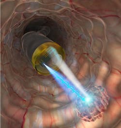
The technique, developed by a team at the Tokyo Institute of Technology, can be implemented using an endoscope that is equipped with spin-LEDs. The method is based on the scattering of circularly polarized light and how it interacts with healthy and unhealthy cells. Specifically, researchers shone near-infrared circularly polarized light on “sliced tissue samples of murine liver containing metastatic lesions derived from intrasplenically injected human pancreatic cancer cells.” They found clear differences in the degree of circular polarization of the light scattered from the samples, depending on the state of the biotissue; this demonstrated that cancer identification is possible with this technique.
“The depolarization of circularly polarized light scattered from biological tissues depends on structural changes in cell nuclei, which can provide valuable information for detecting cancer concealed in healthy tissues,” says Nozomi Nishizawa, an assistant professor in the Tokyo Institute’s Division of Materials Integration, who led the study.
Existing endoscopic diagnosis techniques, such as narrowband imaging, have only been able to confirm the presence of cancer and distinguish between tumorous and nontumorous tissue. According to the Tokyo Institute study, there are very few direct measurement techniques that can provide “a quantitative diagnosis of the depth and area of a carcinoma.”
The new technique could potentially be used for the diagnosis of ulcerative colitis and alcoholic cirrhosis, as well as the observation of engraftments in regenerative medicine and transplant surgery. Reference: N. Nishizawa et al., J. Biophoton. (2020); doi.org/10.1002/jbio.202000380.
About the Author
Justine Murphy
Multimedia Director, Digital Infrastructure
Justine Murphy is the multimedia director for Endeavor Business Media's Digital Infrastructure Group. She is a multiple award-winning writer and editor with more 20 years of experience in newspaper publishing as well as public relations, marketing, and communications. For nearly 10 years, she has covered all facets of the optics and photonics industry as an editor, writer, web news anchor, and podcast host for an internationally reaching magazine publishing company. Her work has earned accolades from the New England Press Association as well as the SIIA/Jesse H. Neal Awards. She received a B.A. from the Massachusetts College of Liberal Arts.

