Combination of advanced imaging techniques brings new applications to light
Over the past decades, researchers have obtained a look at life on many scales and across the spectrum. This has been enabled by the development of a wide variety of fluorescent proteins. By reporting application-specific information through fluorescent light, fluorescent proteins have spawned new fields of biological research. At the same time, improvements in image sensors, illumination sources, and optics have enabled biomedical researchers to get a closer and more detailed look at their samples.
In fluorescence imaging, the light intensity can be quite low, depending on the specific application. Image sensors optimized for low-light conditions, such as electron multiplying charge-coupled devices (EMCCDs) or scientific complementary metal-oxide semiconductor (sCMOS) image sensors, provide the imaging capabilities required for imaging low-light samples. While their light sensitivity and noise characteristics are much better than conventional CMOS image sensors, their image acquisition speeds of a couple hundred frames per second are no match for the high-speed CMOS image sensors that can image at tens of thousands of frames per second.
This presents a tradeoff between light sensitivity and acquisition frame rate. However, some applications involve dynamic phenomena in biological samples that emit little light. These applications require an imaging system that is not only sensitive enough to record the little light available, but is also fast enough to prevent motion blur in the images.
A clever combination of advanced technology opens the door to low-light, super slow-motion imaging. By combining a high-speed image sensor with an image intensifier, low-light imaging can be done at much higher frame rates than current sCMOS image sensors provide. The image intensifier boosts the incoming light to a level that can be imaged via fast CMOS sensors by converting the incoming light to the charge domain, where it is multiplied and then converted back to light. The intensified light is then projected onto a fast CMOS image sensor. This combination of technologies provided by intensified high-speed cameras can result in an increase in light sensitivity of up to two orders of magnitude compared to sCMOS image sensors, and it enables new applications in low-light imaging at high frame rates.
Microfluidics
One example is microfluidic research, where the subject of interest can be a quick-moving droplet of micron scale that emits a very limited amount of fluorescence light. Researchers at the Max Planck Institute (Heidelberg, Germany) developed an alternative method for the functionalization of microfluidic droplets by self-assembly of cholesterol-tagged DNA at the droplet periphery.1 With the droplet generation taking place in just 3 ms, recording this process in any detail requires a frame rate of thousands of frames per second.
However, the amount of light emitted by the sample is limited by the amount of fluorescently labeled DNA in the droplets. The fluorescent signal is not strong enough to be imaged with a conventional high-speed camera. So, the solution is to amplify the emitted light to a level that can be detected by a high-speed image sensor. With an intensified high-speed camera, the researchers were able to capture the droplet formation process in a series of images, which illustrates the generation of one single droplet. In addition, they were able to show that cholesterol-tagged DNA concentrates at the periphery of the droplet.
Cardiovascular and neuroscience research
Other fields where intensified high-speed cameras show their strength are cardiovascular and neuroscience research. In these fields, model organisms such as zebrafish and C. elegans worms are often used to study biological systems on a smaller scale. Discoveries made in model organisms translate well to other organisms, including humans. And because young zebrafish are transparent, their internal organs can be imaged in vivo to see how they function under natural circumstances.
A group of researchers at Columbia University (New York, NY) developed a new microscopy technique called swept confocally aligned planar excitation (SCAPE) microscopy to perform high-speed 3D microscopy in model organisms.2 They applied this technique to record 3D video of a beating zebrafish heart. Generally, the zebrafish heart needs to be paralyzed during imaging to prevent motion blur caused by its beating movements. The HiCAM Fluo, an intensified high-speed camera by Lambert Instruments (Groningen, Netherlands), is fast and sensitive enough to allow the researchers to image a beating zebrafish heart in three dimensions at 321 volumes per second.
To achieve this, the sample is irradiated with light-sheet illumination to image a single plane at a time. The light sheet is swept across the sample to record a stack of images, each of a different plane. By stacking them, a 3D image of the sample is formed. Depending on the number of planes and the image dimensions in each, this method can record hundreds of 3D volumes per second. The resulting 3D videos show the beating zebrafish heart as never seen before—individual red blood cells can be followed as the heart pumps them around. At the same time, waves of green GCaMP spread from the atrium to the ventricle, illustrating the calcium dynamics in the heart.
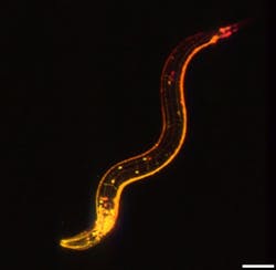
The same microscopy and imaging techniques were applied to perform high-throughput structural imaging in a range of fixed, cleared, and expanded tissues of a freely moving C. elegans worm. Imaging this tiny worm in vivo offers insight into dendritic processes and motion-related neuronal activity at impressive spatial and temporal resolution.This combination of advanced microscopy and imaging techniques enables volumetric imaging at extraordinary speeds. The resulting 3D movies add a new dimension to fluorescence imaging, as they’re able to track movements and dynamic changes in functional fluorescence signals with high precision.
Big data
During the acquisition of the image data for the applications discussed here, a large amount of image data is recorded. The data throughput of such a system is several gigabytes per second, which necessitates a large volume of ultrafast storage.
In the past, high-speed cameras would use in-camera memory to store the image data, but the size of the built-in memory would then limit the recording duration to anywhere between a couple of seconds to half a minute. As data transfer interfaces and computer storage have become faster, it is now possible to stream the image data to the computer and store it in real time. This enables the acquisition of much larger datasets, as the recording duration now scales with the size of the computer storage, which results in recording times in the order of hours, depending on the configuration.
Future developments
The combination of high-speed image sensors and image intensifiers enables new applications that require ultrafast imaging in challenging low-light situations. Together with the improvements in the throughput speed of computer interfaces and storage, new applications that require the acquisition of large amounts of image data are now possible.
New developments of the technologies involved are expected to further improve the light sensitivity of the image sensors and image intensifiers to enable applications where even less light is available. At the same time, further increases in image data acquisition and throughput speeds will enable advanced imaging applications that require more image data.
REFERENCES
1. K. Jahnke et al., Adv. Funct. Mater., 29, 23 (2019); https://doi.org/10.1002/adfm.201808647.
2. V. Venkatakaushik et al., Nat. Methods (2019); https://doi.org/10.1038/s41592-019-0579-4.
Jeroen Wehmeijer | Business Development Manager, Lambert Instruments
Jeroen Wehmeijer is the business development manager at Lambert Instruments (Groningen, Netherlands).
![FIGURE 1. The last eight images out of a series of 12 [1]. Generation of one single droplet (0.003 s) containing Cy3-labeled DNA, captured with a frame rate of 4086 frames per second. FIGURE 1. The last eight images out of a series of 12 [1]. Generation of one single droplet (0.003 s) containing Cy3-labeled DNA, captured with a frame rate of 4086 frames per second.](https://img.laserfocusworld.com/files/base/ebm/lfw/image/2021/09/2110LFW_bow_weh_1.613b6d9cc4120.png?auto=format,compress&fit=max&q=45&w=250&width=250)
