Under the sea: Holographic microscopy gets to the heart of plankton
Plankton is one of the most important creatures in the ocean. It’s the foundation of marine food pyramids—providing food for larger plants and animals, as well as indirectly to humans via fisheries that rely on it.
“Their life has a major impact on our life,” says Harshith Bachimanchi, a doctoral student in the physics department at the University of Gothenburg (Sweden), noting that phytoplankton (microscopic plant-like organisms) produce half of the oxygen we breathe. The number of plankton in the sea impacts the ocean’s entire carbon cycle; collectively, single-cell microplankton use about three times as much carbon as humans emit from fossil fuels.
“But we know surprisingly little about them,” he adds, mainly because of their microscopic size. With larger organisms such as animals or birds, there is a good, solid understanding of who eats who, where the material ends up, how much is lost, etc.
Alongside physics and marine science researchers from Gothenburg, Bachimanchi is now investigating how to alleviate the lack of knowledge with a new microscopy-based technique.
“We set out to obtain individual resolution for microbes to gain a better understanding of the plankton, the most important but least known creature in the ocean,” Bachimanchi says.
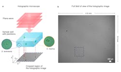
The team’s new approach involves holographic microscopy—this is digital holography, the acquisition and processing of holograms with a digital sensor array, applied to microscopy. Specifically, the researchers use an inline holographic microscope in a lens-less configuration in which the sample containing the plankton is illuminated with a monochromatic LED light source.
The microscope’s sensor is placed below the bottom of the well, allowing the area to be imaged entirely within a single field of view. This way, all of the plankton are continuously visible for the duration of the experiment.
The study stems from work presented several years ago by Giovanni Volpe, a physics professor at Gothenburg who is a co-author of this new study, about the holographic microscopy method. With it, a hologram is created by light that’s refracted through a particle; this allows the hologram to be studied, rather than the particle.
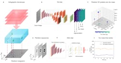
As the light passes through the cells, Bachimanchi explains, it acquires a complex amplitude that depends on the optical properties of the material it traverses. This generates inline holograms or interference patterns on the camera. The holograms encode the 3D position of the plankton as well as the size and refractive index information.
In the Gothenburg study, the plankton was placed under the holographic microscope in two different configurations. For short-time-scale experiments, the microplankton’s dry mass— essentially the mass of a biological sample after the water content has been removed—is estimated and its feeding observed. For the long-time-scale experiments, such as growth and cell division, the diatoms were kept in enclosed circular wells.
The resulting images of the plankton are diffraction patterns formed by the interference of the unscattered light and the light scattered by the plankton.
To analyze the holograms, the team applied a deep learning-based approach that uses two artificial neural networks in sequence. The first—a regression network, or RU-Net—takes the recorded holographic image from the camera and detects the plankton positions along with the dry mass and vertical distances. The holograms (diffraction patterns) act as a unique fingerprint of the size, refractive index, dry mass, and the lateral and axial position of the plankton. “This information is useful to classify planktons by their species, as well,” Bachimanchi says.
The second neural network—weighted-average convolutional network, or WAC-Net—then takes the smaller crops of planktons and refines the information to predict a more accurate value for the dry mass and vertical distance. The microplankton examined with the new technique are no larger than a few hundredths of a millimeter. Traditionally for ecologically relevant microorganisms such as plankton, this small size has meant researchers had to depend on bulk measurements and ensemble averages. Seeing and analyzing individual microplankton with existing microscopy and imaging techniques has basically not been possible.
Another existing challenge with analyzing holographic data is that it’s computationally expensive, Bachimanchi says, especially for a lens-less holographic microscope like the one his team uses in their study. It offers a large field-of-view image that can record more than 700 plankton at the same time, but “the computational processing become enormous,” he adds. “And extracting the dry mass information from inline holograms hasn’t been straightforward.”
The deep-learning algorithms circumvent the computationally intensive processing of holographic data, the study notes, and allows rapid measurements over extended time periods.
The combination of holographic microscopy with deep learning makes the technology more versatile and many order of magnitude faster, which is key to following and characterizing individual plankton throughout their life span. The new approach also provides a strong complementary tool in marine microbial ecology, Bachimanchi says, as it enables a nondestructive and minimally invasive determination of the 3D position and dry mass of individual microorganisms.
“By combining holographic microscopy with artificial intelligence we can follow multiple microorganisms such as plankton and diatoms at the individual level throughout their life span,” Bachimanchi says. “And we can continuously see what they are up to, what they eat, how fast they grow and swim, and so on.”
In addition to working with microbes (microscopic plankton and bacteria, for example)—tracking their movements and whereabouts for extended time periods—the new holographic microscopy/deep learning technique serves as a rapid method for counting, sizing, and weighing cells in suspension. The hardware of the technique is also relatively cheap, Bachimanchi says, and researchers can share their code with examples and demonstrations to facilitate use by other groups.
“Since the microscopic organisms in the ocean constitute the base of the ocean food web and drive large-scale processes at the global scale, the new method permits us to better understand the effect of tiny organisms on larger-scale processes such as the global carbon cycle,” Bachimanchi says. “Our method enables us to follow individual organisms in this soup of life.”

Justine Murphy | Multimedia Director, Digital Infrastructure
Justine Murphy is the multimedia director for Endeavor Business Media's Digital Infrastructure Group. She is a multiple award-winning writer and editor with more 20 years of experience in newspaper publishing as well as public relations, marketing, and communications. For nearly 10 years, she has covered all facets of the optics and photonics industry as an editor, writer, web news anchor, and podcast host for an internationally reaching magazine publishing company. Her work has earned accolades from the New England Press Association as well as the SIIA/Jesse H. Neal Awards. She received a B.A. from the Massachusetts College of Liberal Arts.


