Next-generation microscope boosts opportunities
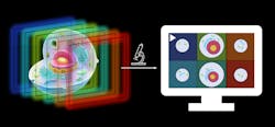
Researchers from UiT Arctic University of Norway and the University Hospital of Northern Norway have developed a multifocus microscope belonging to the fluorescence microscope family. It produces extremely clear 3D images in a natural environment of cell samples tagged with markers that absorb blue light and in turn release green light, and then sorts the images into layers that can be studied from all angles (see video).
Researchers can take images of multiple Z planes during single camera exposure; the team has been able to capture around 100 complete images per second of cells (see Fig. 1). They expect to increase this number with further development.
Multiple Z planes get onto the camera through a cascade of beamsplitters, where each split-off path directs light from the sample plane onto a different region on the camera. Each path has a slightly different yet exactly calculated length and thus images a different Z plane.
During image acquisition, background blur is removed. This is done by changing the illumination from that which floods the entire image to an ultrafast scan of a thin sheet of laser light. At every instance in time, the sheet’s position is conjugated to a specific row on the camera.
“This allows us to trigger the camera’s rolling shutter exposure at just the right time, such that it captures only the light-sheet-illuminated parts of the sample volume, while all other pixels do not record any light, which would otherwise be the blur in the final image,” says Florian Ströhl, a researcher in the Department of Physics and Technology at UiT and project leader of light-sheet microscopy.
After a full light-sheet sweep, the camera is done exposing its frame, which now contains multiple separate Z planes without background haze (see Fig. 2). “Conceptually, this next-generation microscope enables an unprecedented combination of 3D image quality and speed,” Ströhl says.Ströhl, a lead researcher on a recent study published in Optica, developed the concept for this next-generation microscope in 2019 and a year later completed a rather rudimentary proof-of-concept system. With a grant from the Research Council of Norway last year, he and fellow researchers moved forward with a prototype.
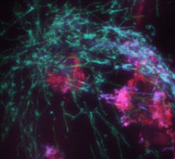
Among applications the microscope has aided is in work by Åsa Birna Birgisdottir, an associate professor in the clinical cardiovascular research group at UiT. She and her team have developed a model system for heart tissue—a mature human heart muscle, cultured completely in a dish from induced pluripotent stem cells—to study mitochondria in heart tissue.
Such cells, which can develop into many different types of cells or tissues in the body, are extremely difficult to take high-resolution pictures, noting that mitochondria (subcellular organelles) are only visible in very-high-resolution microscopes. At the same time, the cells that contain these mitochondria are part of an orders-of-magnitude larger tissue slab, which is highly scattering.
“Taken together, the combined requirement of high-speed 3D imaging with the huge background from the tissue itself basically ruled out being able to use any conventional method,” Ströhl says (see Fig. 3). “Our microscope managed to tick all boxes and could take extraordinarily crisp image volumes.”The plan is to expand on the microscope’s capabilities for other groups to image a variety of model organisms. “With a bit of luck, it will even become a product one can simply buy in the future.” Ströhl says.
About the Author
Justine Murphy
Multimedia Director, Digital Infrastructure
Justine Murphy is the multimedia director for Endeavor Business Media's Digital Infrastructure Group. She is a multiple award-winning writer and editor with more 20 years of experience in newspaper publishing as well as public relations, marketing, and communications. For nearly 10 years, she has covered all facets of the optics and photonics industry as an editor, writer, web news anchor, and podcast host for an internationally reaching magazine publishing company. Her work has earned accolades from the New England Press Association as well as the SIIA/Jesse H. Neal Awards. She received a B.A. from the Massachusetts College of Liberal Arts.


