Innovative photoreceivers simplify measurements
CHARLIE HULTGREN
Semiconductor photodiodes combined with creative analog circuitry are increasingly useful for optical detection applications such as atmospheric, biological, and spectroscopic studies, in which large conversion gain, low noise, and rugged, compact packaging are often critical factors. Femtowatt photoreceivers and balanced photoreceivers are examples of two such devices.
In creating a femtowatt photoreceiver, the photodiode’s sensitivity can be enhanced by combining it with a simple operational-amplifier/resistive feedback circuit. Because the feedback resistor must be large to achieve high gain, these circuits often suffer from significant amounts of Johnson noise. Careful choice of the amplifier-resistor pair, however, allows one to build detectors with 2 × 10¹¹ V/W gain and noise equivalent power of less than 16 fW/Hz. In some applications, these detector-circuit combinations can be used as simple, battery-powered substitutes for photomultiplier tubes.
The balanced photoreceiver is another instrument that allows sensitive measurements. This kind of photoreceiver is ideal in any experimental setup that has reference and signal beams. Differential techniques cancel out laser-intensity noise or any other common-mode noise. An auto-balanced photoreceiver we developed at New Focus (Santa Clara, CA) is based on two silicon or indium gallium arsenide (InGaAs) photodiodes and a differential amplifier. The two photodiodes, one each for the signal and reference beams, are followed by a patented auto-balancing circuit that subtracts the two photocurrents and cancels noise signals common to both channels. In addition, the auto-balancing circuit automatically produces a balanced output, even if the average optical intensities on the two detectors are different and time-varying. This allows detection of the weak signals commonly found in laser spectroscopy applications. This device also enables reduction of common-mode noise by more than 50 dB at frequencies from dc to 125 kHz—effectively eliminating laser intensity noise and allowing shot-noise-limited measurements at low frequencies without lock-in amplifiers and optical choppers.
Three examples illustrate the usefulness of these specialty receivers. The first two take advantage of the photodiode/ high-gain amplifier—one for studying the upper mesosphere, the other for studying calcium ions in biological cells. The third example describes a balanced photoreceiver used in a rubidium spectroscopy application.
Atmospheric potassium
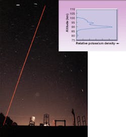
The potassium resonance lidar at the Arecibo Observatory (Arecibo, Puerto Rico) is used to study dynamics and chemistry in the upper mesosphere. Resonance lidar takes advantage of meteor-deposited metals such as sodium (Na) and potassium (K) in the upper mesosphere (80–110 km above the ground) of the earth’s atmosphere (see photo at top of this page). The coldest region of the atmosphere, the mesopause, normally occurs at this altitude, making it a region of intense geophysical and climatological interest.
The beauty of having an atomic metal layer in this rarefied region of the atmosphere is that the spectra of atomic metals are well understood and high-resolution spectroscopic measurement of this remotely situated sample can provide insight into the atmospheric conditions surrounding the sample. The spectral lineshape, for example, is sensitive to the temperature. In addition, once a local frequency marker has been obtained for purposes of comparison, the Doppler shift of the spectrum can give the line-of-sight wind speed.
The femtowatt photoreceiver plays a critical role in obtaining the local frequency marker. Our de vice uses a standard silicon or InGaAs photodiode followed by a transimpedance amplifier with extremely high gain settings (2 × 10¹⁰ and 2 × 10¹¹ V/A). This high-gain amplifier is optimized for low-noise performance, enabling detection of weak optical signals at levels of less than one nanowatt down to a few femtowatts. A pulsed-output alexandrite laser is used for the lidar transmitter and, in order to excite the mesospheric potassium, its output must be locked to the potassium resonance line with extremely narrow spectral linewidth and high precision. The spectral locking and linewidth are maintained by a continuous-wave (CW) injection seed laser—in this case an external-cavity diode laser.
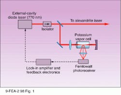
The seed laser is locked to the Doppler-free feature of the K-D1 resonance at 770 nm (see Fig. 1). A small amount (less than 1%) of the seed laser light is transmitted through a cell containing pure potassium vapor. A mirror on the opposite side of the cell retroreflects the laser so that there are two counterpropagating beams in the cell. Once the laser is tuned to the D1 resonance, the potassium fluoresces. If the laser is not tuned to the exact center of the line, the beam interacts with atoms whose motion along the beam direction creates a Doppler-based shift of the laser frequency onto resonance. Thus, each of the counterpropagating beams interacts with a different population of atoms. But when tuned to the line center, each beam interacts with the same population (zero velocity) of atoms, which results in a saturation of the interaction and a dip in the fluorescence intensity.
A femtowatt photoreceiver operating in the lower-gain dc mode (2 × 10¹⁰ V/A) detects the fluorescence. The output from the receiver is fed to a lock-box that consists of a lock-in amplifier and a modulator. The modulator and lock-in output modulate and maintain the laser wavelength on the Doppler-free feature. Once locked by this method, the remainder of the seed laser output is used to injection-seed the alexandrite laser. The alexandrite beam is transmitted to the sky, and the return scattering from the mesopause is detected through an 800-mm Cassegrain telescope coupled to a photomultiplier, yielding a K-profile based on a one-minute integration with 150-m altitude resolution (see inset to photo at top of this page). Note that the signal comes from a narrow range of 80–100 km. More on K-resonance lidar has been published by other groups.1, 2
Finally, by strategically choosing three points on the K-spectrum and tuning the laser alternately to each of them, temperature and line-of-sight wind speeds in the mesosphere can be measured. This frequency switching is done by locking the seed laser, then frequency modulating the output with a pair of acousto-optic modulators in a unique arrangement.3
Biological fluorescence
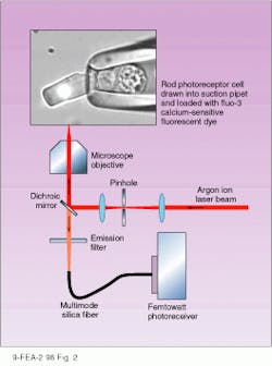
Another application of the femtowatt photoreceiver involves fluorescence detection to measure calcium ion concentrations within single living photoreceptor cells from the human retina. The photoreceiver is used in a laser spot confocal technique to measure the fluorescence emitted from a calcium-sensitive probe molecule incorporated into single rod and cone photoreceptors (see Fig. 2).
An argon-ion laser emitting at 514.5 nm illuminates a 50-µm-diameter pinhole. Then a 4X microscope objective recollimates the pinhole light before it passes into the epifluorescence port of a Nikon Eclipse 300 inverted microscope. A dichroic mirror reflects the beam, and a high-numerical-aperture 40X oil immersion lens images the beam onto the outer segment of a single living photoreceptor cell—loaded with the calcium-sensitive fluorescent dye fluo-3—to yield a 5-µm fluorescence-exciting spot. The fluorescence excited by the laser spot is collected by the oil immersion lens, passed through the dichroic mirror and an emission filter, and imaged onto a 200-µm multimode optical fiber coupled to the femtowatt photoreceiver.
Because the fiber coupled to the detector is matched in size to the spot image (5 µm × 40X magnification = 200 µm), this system is truly confocal, collecting fluorescence selectively from the volume element selected, which is critical to avoiding spurious fluorescence signals from unwanted regions of the cell.
A previous version of this experiment used a small-area photodiode placed directly in the image plane and coupled to an integrating current-to-voltage converter designed for low-level biological current measurement. The drawback of that approach was the high cost of the current-to-voltage converter. Also, the direct placement of the detector in the image plane made interchange of detectors difficult. The current femtowatt photoreceiver is small, relatively inexpensive, and almost as quiet as the previous detector/amplifier combination. Further, when fiber-coupled, this setup provides the robustness of a semiconductor photodiode with the flexibility of using other detectors for even lower-light measurements.
This experiment aims to determine the role of cytoplasmic calcium concentration in retinal photoreceptor cells. According to widely held scientific opinion, a decrease in calcium concentration may cause the changes in photoreceptor sensitivity that occur during exposure to light. The decrease in calcium concentration may also be implicated in diseases that cause the retina to degenerate. Researchers at the University of Cambridge (Cambridge, England) plan to use this technique to investigate stimulus-induced changes in cytoplasmic calcium concentration in olfactory receptor cells from the nasal epithelium, which are responsible for the sense of smell.
Another specialty photoreceiver, the auto-balanced photoreceiver, can reduce noise sources that plague many precision spectroscopy measurements. The device is ideal for use in the type of dual-beam setup commonly found in atomic and molecular spectroscopy—one invariant reference path and another path containing the signal of interest. In this case, the circuitry subtracts the reference and signal photocurrents, canceling noise signals common to both channels.
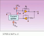
The auto-balancing circuit uses a low-frequency feedback loop to maintain automatic dc balance between the signal and reference arms. The circuit consists of two photodiodes, a current splitter, a current subtraction node, a transimpedance amplifier, and a feedback amplifier (see Fig. 3). In auto-balanced mode, the current-splitter gain g is electronically controlled by a low-frequency feedback loop to maintain equal dc photocurrents. In this mode the receiver is automatically balanced, canceling laser noise regardless of the photocurrent.
In a test of the noise-reduction capability of the device, a tunable diode laser was used to perform an absorption measurement of a rubidium line. At the output of the diode laser an amplitude modulator added noise to the laser signal for purposes of the demonstration. Then, the laser was split into a reference and a signal beam, and the signal beam was sent through an atomic rubidium cell. With the device operating in auto-balanced mode, much of the noise obtained with a standard absorption measurement technique using just one photodetector is canceled.
ACKNOWLEDGMENTS
Jonathan Friedman provided data and a description of the atmospheric potassium density measurements performed at Arecibo Observatory. Paul Castleberg obtained the experimental data shown in the inset to the photo at the top of this page. The Arecibo Observatory is operated by Cornell University under a cooperative agreement with the National Science Foundation.
Hugh R. Matthews at the University of Cambridge provided a description of the laser spot confocal measurement technique. The work described was performed by Matthews and Johannes Reisert, Physiological Laboratory, University of Cambridge; Alapakkam P. Sampath (Department of Physiological Science) and Gordon L. Fain (Departments of Physiological Science and Ophthalmology), University of California, Los Angeles; and M. Carter Cornwall, Department of Physiology, Boston University Medical School.4,5
The spot confocal measurement technique was originally developed for studies of skeletal muscle by Julio Vergara of UCLA; the approach described here was derived from his work.6
REFERENCES
1. G. Megie et al., Planet. Space Sci. 26, 27 (1978).
2. U. Von Zahn and J. Höffner, Geophys. Res. Letters 23, 141 (1996).
3. C. Y. She and J. R. Yu, Geophys. Res. Lett. 21, 1771 (1994).
4. H. R. Matthews et al., J. Physiology-London 506P, 3 (1998).
5. A. P. Sampath et al., J. General Physiology 111, 53 (1998).
6. A. L. Escobar et al., Nature 367, 739 (1994).


