MEDICAL IMAGING: Fiber delivery systems enhance flow cytometry designs
FIONA EVANS
Advanced research in gene expression and the growing market for high-throughput clinical and diagnostic screening are driving step changes in biophotonics technology. These advances enable the applications to leave the arena of academic research and be used in high-volume industrial-scale research, with the ultimate aim of bringing personalized genomics and thereby personalized medicine ever closer.
Many of the screening instruments use high-specification lasers, advanced optical systems, and detectors to achieve reliable, high-speed, high-resolution analysis of very small target samples in microfluidics or microarrays. Analytical processes such as DNA and blood analysis—both enabled by flow cytometry techniques—rely upon optical system designs, which in turn are based on free-space optics or optical fiber delivery systems.
DNA and blood analysis
DNA analysis is, after immunofluorescence, the second most important application of flow cytometry and it has driven advances in flow cytometry instrumentation. The first mapping of the human genome took ten years to complete; now it takes less than three days, although the data analysis takes much longer. Information can also now be gained more quickly from shorter partial DNA and RNA read lengths to investigate target sections of the DNA strand in order to answer specific queries, such as those related to hereditary diseases or cell and gene mutations as cancer indicators.
Working with what are often irreplaceable samples, researchers in this field need instrumentation that can reliably gather data in a short time, and in a manner that is repeatable between samples and over the long term. The stability and resolution of the instrument is critical and any changes will have a significant impact on the quality of the data and the results.
In a similar fashion, blood analysis for diagnostic and immunology purposes is a large and rapidly expanding market. Advances in genetic research are now leading to new diagnostic tests and test equipment.
The design of all these analytical instruments is as much about reducing sources of error as it is about improving performance, and optical fiber delivery of the lasers is a useful tool that addresses both concerns. Optical fiber delivers the additional benefit of allowing the instrument designer to place the lasers in an easily accessible location for field servicing.
Analysis techniques
The instruments used in these new fields of gene expression and pharmacogenomics are based on the principles of microscopy and flow cytometry. Speed and throughput are key drivers in the design of these instruments, which operate on a busy daily schedule in core facilities to accommodate a large and growing demand for analysis.
Some of these facilities are unable to increase output without building additional laboratory space. Consequently, there is a need for smaller-footprint instruments so the core facilities can fit more instruments into existing laboratories.
In flow cytometry, each excitation laser source usually is dedicated to one probe on the sample per run. One way to speed up the analysis is to run parallel processes by using multiple illumination wavelengths fired in series on the sample. The primary challenge is to focus the light onto a moving sample in the flow stream, which is usually less than 100 μm wide.
To accrue meaningful data from a moving target, both the detector and illumination source need to be as stable and stationary as possible; otherwise, movement from either one will cause image jitter and reduce resolution. The second physical challenge is often positioning the beams from multiple light sources in sufficient proximity to generate parallel illumination spots focused in the flow cell, but also to sufficiently separate these spots to prevent crosstalk in the detection channels, which may collect emitted and fluorescent light simultaneously.
In addition, in genetic analysis the instruments are working with much smaller sample sizes and some of them now image at the single-molecule level. This means that the challenges originally faced by flow cytometry have increased.
Free-space optical systems
Flow cytometers have traditionally used an optical system of lenses, filters, and microscope objectives to shape the beam and deliver it to the flow cell. The principles are well understood, although this often results in a long optical beam train, making it more susceptible to the effects of laser jitter. An open beam path with optics mounted at various locations is also more susceptible to thermal temperature differences that will cause beam movement on the sample.
Careful alignment of the optics is necessary as small movements can cause large changes in the beam pointing on the sample. As a result, instruments that use free-space optics can be susceptible to physical knocks and changes in environmental conditions, including heat generated by the laser.
Nevertheless, free-space optics are still widely used in routine flow cytometer applications. The best designs have been refined to keep the optical path as short as possible, and to use additional cooling methods to regulate the temperature inside the housing.
Fiber-optic delivery
For demanding applications such as microbial screening and gene expression, the use of singlemode fiber to deliver the laser beam to the sample offers several advantages in functionality, from increased laser stability and image resolution to reduced instrument size and greater ruggedness.
Using singlemode fiber dramatically reduces the effect of laser beam-pointing instability (or jitter) regardless of the laser technology employed. In the best systems, the improvement can range from 30 μrad/°C to better than 1 μrad/°C. The fiber also acts as a spatial filter and eliminates beam astigmatism to create a near-perfect Gaussian profile. The resulting beam is much more stable over time than those in free-space systems, and is without accumulated errors from multiple optical interfaces, which ensures a more reliable and more stable measurement (see Fig. 1).
Enclosed in the fiber, the beam is much less sensitive to movement and thermal temperature changes, so this reduces the need for instrument alignment service visits and creates a more robust instrument design. Fiber also allows the laser source, which generates heat, to be located away from the flow cell, which means more flexibility for instrument layout, including the option of mounting the laser externally to the instrument head. It also makes servicing considerably easier, since the optical alignment of the instrument is fixed and independent of the laser, so there is no need to perform a lengthy re-alignment process when the laser is serviced.
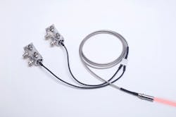
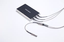
To meet the need for a smaller beam path to reduce instrument footprint and still deliver multiple illumination spots focused in a flow cell, Qioptiq created a singlemode polarization-aligned fiber array, the kineFLEX-Hydra, which delivers two or more spatially separated and parallel beams to the flow cell (see Fig. 2).Each fiber core is actively aligned with submicron precision to achieve the required beam-polarization axis parallelism. Beam-shaping optics and filters can also be integrated into the fiber array to further reduce the optical train. In collaboration with one instrument manufacturer, this approach enabled a two times reduction in footprint.
This development is significant because the result is a robust measurement head, in which all the required beam-shaping and beam-delivery optics are enclosed. Essentially, a complex optical system, which would until very recently have required complex optical setup and alignment, has been reduced to a plug-in component less than 200 mm long (see Fig. 3).Customized applications
Qioptiq has worked alongside researchers and original equipment suppliers in this field to develop instrumentation that enables new possibilities in analysis. For example, one genome screening system now available is able to scan biological samples to detect and identify strains of infectious organisms (e.g., E .coli, Staphylococcus) in less than three hours; in the past, this process would have taken a number of days. This significantly reduces the time between diagnosis and treatment, enabling the targeted use of anti-infectives for optimal effect.
The fiber optic deliver system with integrated beam shaping overcomes the engineering challenge of producing multiple parallel and polarization-aligned beams with tightly controlled spacing in a robust small package.
Qioptiq has also partnered with an equipment manufacturer to provide improved laser stability with a custom solution that enables a five times improvement in measurement resolution in their latest microarray scanner for gene expression. The smaller the target sample, the greater amount of control is required from the illumination source.
Fiona Evans is business development manager at Qioptiq, Mitchell Point, Ensign Way, Hamble, UK S031 4RF; e-mail: [email protected]; www.qioptiq.com.


