A cranioplastic surgeon, working before a holographic image of a patient`s skull, carefully molds clay to conform exactly to the image of the skull. As soon as the front is finished, the image is flipped 180° about its x-axis for anatomical review from the reverse orientation. The finished product—essentially a plastic bone—will be inserted into the patient`s head to replace a damaged portion of the skull. The holographic image is a Voxgram, made with a digital holography system from Voxel (Laguna Hills, CA).
"A surgeon is more a sculptor than a painter," says William Bergman, Associate Chief of Neurosurgery and Director of Neurosurgical Research at Santa Clara Valley Medical Center (San Jose, CA). "We don`t deal with flat pictures. We`ve come to appreciate two-dimensional anatomy from x-rays, but it is more realistic to work with life-like images."
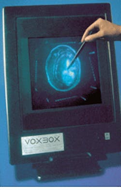
The Voxgram image supplies just that—a life-size, transparent hologram of some part of the body that extends out into space. A physician can interact in, around, and through this image as if it were a real specimen of anatomy (see photo at right). Bergman likens it to a familiar childhood toy—the Visible Man or Woman. "It had a clear plastic body with removable organs, tissues, and bones," says Bergman. "The Voxgrams are just like that. They are transparent images made from light. One can put an instrument inside the picture and see where it is going and what it is going to do."
Holograms with a twist
A Voxgram is a hologram with a twist. Traditional holog- raphy requires that light be reflected off a real object. Because it is difficult to project light inside a body, the "object" for a Voxgram begins with digital data collected from a computed tomography (CT) or magnetic resonance (MR) scan. A CT or MR scan acquires a series of two-dimensional (2-D) pictures or "slices" of a person`s body using x-rays. Typically, these images are viewed as sequential slices. Occasionally they are reformatted into views from other projections or as a 3-D rendering using standard software techniques.
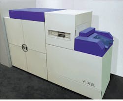
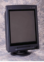
To create a Voxgram—a bright, distortion-free, true three-dimensional image—these tomographic gray-scale slices are projected on a screen at specific distances from the holographic film plane inside a special camera, the Voxcam (see Fig. 1). As the first slice is projected, and its holographic interference pattern is recorded on film using a Coherent (Santa Clara, CA) Compass laser. This light source is a 100-mW frequency-doubled diode-pumped Nd:YAG laser operating at 532 nm. The screen is repositioned to correspond to the gap between the slices. The next slice is projected, and its interference pattern is superimposed on the first. The process is repeated for subsequent slices. Raymond Schulz, director of marketing for Voxel, says that the process is similar to a multiple-exposure photograph when two exposures are made on a single piece of film. The multiple-exposure hologram, however, encodes the depths as well as the intensities for exposure. When reconstructed, all the slices appear "hanging" in space with the correct separation between them. One multiple-exposure hologram may contain more than 200 exposures.The light emitted from the Voxbox lightbox interacts with the interference pattern stored in the holographic film emulsion. The resultant hologram features the same high-resolution volumetric (full aperture) display that laser illumination produces. Voxgrams can be viewed within a conical viewing volume of approximately 45°, meaning they can be viewed vertically as well as horizontally. Because they are volumetric transmission holograms, they can also be flipped over to reveal the anatomy from the reverse side.
On-site imaging
Bergman started using the Voxgram as a model for pre-operative planning. "But then I took it into the operating room," he says. "When you are in the middle of surgery, you want to be spoon-fed information. You don`t want to take the added time to figure something out." In addition to the cranioplastic application he developed for sculpting replacement bone, Bergman combines digital holography with stereotactic technology to "see" into areas that are not easily visualized.
In neurosurgery, one needs to appreciate the exact location in an opaque sphere, such as the head, says Bergman. A surgeon drills a hole and inserts a cannula through to a certain spot. "You have two points—the hole you drilled and the target you are trying to hit. The line in between the two points is what you have to try to follow. But the stereotactic information doesn`t tell you anything about what is in between. Using a Voxgram, I can see everything I am going to hit," he says. Bergman`s work was most recently presented at two separate meetings in October, at this year`s Congress of Neurological Surgeons meeting in New Orleans, LA, and at the North American Spine Society meeting New York City.
Doctors are also using digital holography to aid in breast imaging, plastic surgery, and atherosclerotic disease. David Furnas, Clinical Professor and Chief of the Division of Plastic Surgery at the University of California-Irvine Medical Center (Irvine, CA), uses Voxgrams to help him reshape congenitally malformed skulls. The practice, cranio-facial surgery, involves patient makeovers with skull deformations caused by premature suture lines of fibrous tissue. For example, a child may suffer from a premature suture between the front and back portions of the skull. The suture forces the bone to bulge on one side causing a keel shape. In such as case, Furnas reshapes the skull by designing a gap. He cuts the deformed portion of the skull into components that are reassembled as a self-supporting cranial framework that gives the brain plenty of room to grow. "I put a hologram on the screen to make patterns and templates that determine my cuts and how I should move the fragments," Furnas says. "The hologram is a life-size transparent skull that I can work directly from. A CT scan is about 1/5th the size because it shows slices only. The Voxgram looks like you should be able to touch it."
Jill Hunter, staff neuroradiologist at Children`s Hospital of Philadelphia (Philadelphia, PA), assessed the applicability of digital holography for displaying congenital anomalies of the cervical spine. "These abnormalities can be extraordinarily hard to interpret from a stack of 2-D axial slices," says Hunter. Holograms made from CT data gathered from five sequential cases of congenital cranio-cervical anomalies were used to pre-plan surgery. The transparent nature of the film and the ability of the holograms to be inverted to view the data set from the opposite direction aided the surgeon`s understanding of the "unexpected relationship of these often abnormally formed bones." An added touch was that the holograms helped the parents understand their child`s problem.
During the past five years, more than 165 peer-reviewed presentations have been given at medical meetings both domestically and internationally by radiologists, physicists, neurosurgeons, orthopedic surgeons, and plastic surgeons touting potential applications for holography. William Bergman was director of neurosurgical research at Walter Reed Army Medical Center (Washington, DC) when someone brought a Voxbox to a seminar and asked, "Okay, what can you do with this?" After 10 successful applications and 19 presentations, Bergman says he is just scratching the surface.
Achieving the third dimension
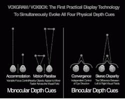
Many computer software programs promise three-dimensional viewing. However, even though an enhanced image appears to have depth or volume, it is still only two-dimensional, primarily because of the nature of the flat-panel display. The human visual system requires both physical and psychological depth cues to appreciate the third dimension. Physical depth cues can be evoked only by true three-dimensional objects; psychological cues can be triggered by two-dimensional pictures (see figure).The four physical depth cues required by the brain to "see" true 3-D are focus, convergence, motion parallax, and stereo disparity. Focus, or accommodation, is the ability of the eyes to focus on nearby objects. This allows us to gauge depth by sensing how much the eye lenses change shape in order to deliver a sharp image of a given object to the brain.
Convergence refers to the distance the eyes have to cross or "toe-in" in order to mentally fuse left-eye and right-eye views. The closer the object, the more the eyes must converge.
Motion parallax provides depth information by comparing the relative motion of different elements of the viewed object. Closer objects appear to move faster across our visual field than those further away.
Stereo disparity refers to the binocular disparity or parallax between left-eye and right-eye images. This is the double image of an object seen when the eyes focus on another object at a different distance. The further away the original object, the further apart are the two images.
Two-dimensional photographic depth information is extracted through four psychological depth cues. These are linear perspective, shading, texture, and prior knowledge. Linear perspective is the perception of the apparent shape of an object relative to its distance from the observer, such as the illusion of railroad tracks converging at a distant point on the horizon. Shading refers to the scattering of light from the surface of an object and is dependent on the angles between the light source, the surface, and the observers. Shadows cast by one object upon another give strong spatial-relationship clues. Variations in intensity help us to infer the surface shape and orientation of an object. Variations and clarity in texture, the small-scale structures on an object`s surface, are interpreted as being due to a change in distance of the surface. The closer the object, the coarser its texture appears to be.
Perhaps the strongest psychological depth cue is prior knowledge. Voxel`s Raymond Schulz explains that we rely on our accumulated knowledge of the common structures of objects—the way light interacts with their surfaces and how they behave when in motion—to first recognize an object. Then we can infer its depth using the other psychological cues. "This is the most important limitation of software methods for producing `pseudo-3-D` medical images," says Schulz. "If we cannot recognize the object, as is often the case if we have not previously seen it, then the psychological depth cues are of little use. They can even lead to a mistaken interpretation." Although psychological depth cues in a 2-D image are powerful, a 2-D image cannot successfully appear as a 3-D image because the four physical depth cues cannot be evoked simultaneously.
Because this text is written on flat, two-dimensional paper, the real volumetric hologram effect is lost. In fact, no slide, photograph, painting, computer screen, or video is capable of displaying true 3-D.
About the Author
Laurie Ann Peach
Assistant Editor, Technology
Laurie Ann Peach was Assistant Editor, Technology at Laser Focus World.


