Confocal Microscopy/Ophthalmology: Towards noninvasive detection of diabetic neuropathy
BÜLENT PEKER
If current trends continue, by the year 2035 one adult in 10 will suffer from diabetes mellitus, a complex disease that is becoming an ever more pressing medical challenge.1 Understanding the mechanisms and implications of diabetes is important for treatment, and researchers at the University of Rostock (Germany) have found confocal laser scanning microscopy to be an ideal tool for this application.
Compared to standard widefield microscopy, confocal laser scanning microscopy delivers a sharp image derived only from a defined focal plane. As confocal technologies have progressed, it has been an interesting development that, while complex modular systems (such as those built on multiphoton technology) enable the most cutting-edge in vivo investigations, a new category of compact all-in-one system has emerged to address everyday demands in life science research laboratories. At the University of Rostock, researchers rely on one such system to study many aspects of diabetes, at the molecular,2 organelle, and cellular levels.
Because not all researchers are familiar with microscopy when they join the group, an easy-to-use system presents many benefits. "We have a lot of students working on their theses," says Simone Baltrusch, vice director of the Institute for Medical Biochemistry and Molecular Biology. "They quickly become independent analyzing their samples with this system [an Olympus FluoView FV10i], which never happened in the past." Ease of use also allows the two postdoctoral researchers in the group to more effectively look after many students: A user-friendly system allows supervisors to be available for help while enabling students to achieve good results independently.
Quantifying hyperglycemia-induced damage
Diabetes can have a huge impact on quality of life, and a common effect is vision loss. One particularly interesting project of the Baltrusch group is quantifying hyperglycemia-induced damage to neuronal networks within the eye. This effort uses confocal microscopy to develop a new technique for early detection of neuronal damage, or diabetic neuropathy.
A key factor underlying diabetes is impaired function of the insulin-producing β-cells of the pancreas, with a subsequent drop in insulin secretion leading to abnormally elevated blood sugar levels (hyperglycemia), which results in a variety of physiological complications. While type 1 diabetes stems from the autoimmune destruction of β-cells, type 2 diabetes is characterized by both β-cell failure and insulin resistance in tissues throughout the body. No matter the type, when hyperglycemia persists over five years or longer, physiological damage can be expansive, and neuropathy is the most common long-term complication. It arises when glycation of blood proteins gives rise to what are known as advanced glycation end products (AGEs). AGEs bind to receptors on the surfaces of neurons, and trigger apoptosis (cell death) and other damaging processes.
Early diagnosis is crucial for disease management, and yet current techniques are nonquantifiable—depending, for instance, on a patient's verbal response to a sensory trigger applied to the foot. While analyzing the neuronal network from a skin biopsy can identify any fibers lost, this is painful, and introduces a complication as wound healing is problematic for people with diabetes. A viable quantitative technique is needed.
Together with students of Prof. Oliver Stachs, group leader of the Department of Ophthalmology at the university, Prof. Baltrusch's team is instead working to analyze neuronal networks in the cornea of the eye. Using a specially designed corneal confocal microscopy system, the ophthalmology team is able to visualize the subbasal nerve plexus of the cornea in humans.3 This noninvasive approach lends itself to the detection of diabetic neuropathy, and the researchers are both understanding more about the structure of corneal nerves and developing this as a quantitative technique within the clinic. Working with diabetic mouse models, confocal microscopy is applied to quantify both nerve density and length within isolated corneas ex vivo, providing insights into diabetic neuropathy and guiding the most effective use of in vivo corneal confocal microscopy.
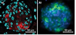
With a focus on a non-autoimmune triggered type of diabetes, researchers specifically destroy the β-cells of the pancreatic islet cells using the compound streptozotocin, investigating samples using antibody staining for insulin, and visualizing them using confocal microscopy (see Fig. 1). Visualizing the corneal nerves of the thy1-YFP transgenic mouse ex vivo, they are able to distinguish the larger stromal nerve from the very thin nerves at the subbasal nerve plexus (see Fig. 2). The cellular architecture of the cornea is complex and only the latter type of neurons are of interest, comments Prof. Baltrusch: "The subbasal nerves are very sensitive to damage, and you see a loss of these in people and animals with diabetes." Comparing old and new techniques, skin samples from the mice are also analyzed, and corneal nerves are found to be much more susceptible-owing to having more receptors for AGEs.While it is crucial to find the right layer of the cornea and analyze the correct nerves, being so thin this can be challenging. Confocal microscopy allows selection of the correct z-position and facilitates analysis with 3D imaging.
A bigger picture
Another indicator of neuronal health is nerve fiber length. Seeing the length of nerve fibers requires a large field of view—which is made possible using automated image stitching. "One of the main reasons why we chose this system is for image quantification and high-throughput analysis," says Prof. Baltrusch. "With the image stitching on this system we get a very nice overview."
Figure 3 shows an acquisition of 36 images with 25 z-slices generated overnight; imaging one quarter of the cornea takes three days. Throughout such extended experiments, the machine runs autonomously. An autofocus routine ensures that the scan field remains in focus between multiple images. Such automation is not standard on confocal setups, especially not with a compact all-in-one system. Without automation, however, an operator would have to focus each image, and need experience to produce a high-quality result.For quantifying nerve fiber density and the length of individual nerves, image stitching yields a much more insightful representation of the cornea. "Ideally we want to look at the whole cornea," says Prof. Baltrusch. The corresponding data load (the image representing less than a quarter of the cornea constitutes approximately 6 GB) requires specialized software for image analysis and quantification.
Follow-on investigations
"Even for those without microscopy experience, we can now analyze approximately 1000 samples throughout all tissues to create a picture of diabetes in the whole organism," comments Prof. Baltrusch. Allowing students and professors alike to generate high-quality results is a key driver for advancing academic research.
In addition to facilitating the detection of neuropathy, these investigations have opened other avenues in research. Prof. Baltrusch goes on to say: "We are interested in which areas are damaged first, and when mice are treated with insulin, what comes back first? What is really interesting is that you can see [in Fig. 3b] these dots in the nerves, and we know that these beaded nerve fibers branch and thus are important for new nerves."
Indeed, combining accurate diabetic mouse models with in vivo and ex vivo confocal microscopy produces an ideal platform not just for diagnosis, but also for testing new compounds that might treat diabetic neuropathy.
ACKNOWLEDGEMENT
Professor Dr. Simone Baltrusch is vice director of the Institute for Medical Biochemistry and Molecular Biology, University of Rostock, Germany. She achieved her PhD from the University of Braunschweig and her venia legendi from the Hannover Medical School, employing microscopy to study protein-protein interactions. Baltrusch has since developed a particular focus on the physiology and pathophysiology of diabetes mellitus. She moved to the University of Rostock for her professorship in 2008 and, in 2009, received the Ernst-Friedrich-Pfeiffer prize from the German Diabetes Association. She can be contacted at [email protected].
REFERENCES
1. See http://bit.ly/1iiBkyH.
2. A. Hofmeister-Brix et al., J. Biol. Chem., 288, 50, 35824–35839 (2013).
3. A. Zhivov et al., PLoS ONE, 8, 1, e52157 (2013); doi:10.1371/journal.pone.0052157.
Bülent Peker is Product Manager of Laser Scanning Microscopy at Olympus Europa (Hamburg, Germany); www.olympus-lifescience.com.


![FIGURE 3. Visualizing the thy1-YFP mouse cornea with image stitching. Multi-area time-lapse viewer shows 36 single images with 25 z-layers each (a). The image in the marked white square is shown as a 3D maximum projection (b), and as a 3D volume view (c). Image stitching facilitates analysis of subbasal corneal nerves (red square in [a], and [d]). FIGURE 3. Visualizing the thy1-YFP mouse cornea with image stitching. Multi-area time-lapse viewer shows 36 single images with 25 z-layers each (a). The image in the marked white square is shown as a 3D maximum projection (b), and as a 3D volume view (c). Image stitching facilitates analysis of subbasal corneal nerves (red square in [a], and [d]).](https://img.laserfocusworld.com/files/base/ebm/lfw/image/2016/01/1507lfw_sch_f3.png?auto=format,compress&fit=max&q=45&w=250&width=250)