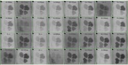
Spectral imaging combines spectroscopy and camera visualization to provide scientists with a more complete understanding of life. The technique captures chemical composition along with spatial structure, and represents these characteristics by way of spectral graphs and images. Varieties of the technology go by such names as hyperspectral imaging, multispectral imaging, imaging spectroscopy, and even chemical sensing. David Bannon, chief executive officer at Headwall Photonics (Fitchburg, MA), says, "It's spectroscopic technology by which someone can actually generate a chemical signature of all of the objects in a scene." To help life scientists explore this technology, Headwall Photonics and others, including the Belgium-based research organization imec, make starter kits.
Value and growth
Spectral imaging can be performed in several ways. The key is that a sample gets scanned visually and across some spectral range. For example, a grating or prism can be used to separate the wavelengths of light, thereby collecting the entire spectrum at once. Explaining the value of this imaging technique, Bannon says: "The key point is capturing all of the spatial and spectral information in the scene, creating a very large data set and searching it for particular chemical signatures while maintaining the spatial information." He adds, "Every object has a unique spectral signature, which is a function of the way the object reflects and absorbs light."
Spectral imaging appears in an increasing number of life science projects. "More and more applications of spectral imaging have been proven to be very interesting in research environments," says Andy Lambrechts, team leader of the hyperspectral imaging R&D team at imec. "People are looking for more economically viable ways to use this kind of imaging." He adds, "New technology is becoming available that removes some of the bottlenecks that prevented people from taking the first step in using this technology."
As this technology becomes more accessible, it will spread throughout the life sciences. "This is like a feedback loop," Lambrechts says. "Increased interest makes more people be aware of this technology, and that is boosting the novel applications. It will pop up everywhere in the coming years."
Driven by discovery
Many kinds of biological advances can propel an interest in spectral imaging. "Two primary developments in the past two decades were knowledge of genomes and very fast and very high-resolution biological imaging," says Gang-yu Liu, professor of chemistry at the University of California at Davis. "This combination, once it reaches molecular resolution, shall enable us to understand the impact of genomics in conjunction with the local environment, the molecular-level environment."
To take on this challenge, Liu teamed up with Alan Hicklin, development engineer at the University of California, Davis. They wanted to create a system that provides high spatial resolution combined with molecular information. For the spatial resolution, they turned to one of Liu's areas of expertise, atomic force microscopy (AFM), in which a nano-size tip scans the surface of a specimen. This provides a topographic map of sorts, revealing features as small as fractions of a nanometer.
For the molecular information, Liu and Hicklin added spectroscopy. "This tells us what kind of molecular or functional groups exist in a sample," Liu explains.
The spectroscopic imaging comes from a scanning confocal microscope or a scanning near-field optical microscope. "It uses several laser lines, from near-infrared to near-ultraviolet, 400 to 750 nm," Hicklin says.
The combined microscopy platform that Liu and Hicklin developed scans a sample with AFM from the top and scanning confocal-spectral microscopy from below. Making these two technologies work together creates challenges of scale. "Atomic force microscopy looks at a few molecules, and the confocal image comes from tens of thousands of molecules," says Hicklin.
To ensure correlation between the topograph and spectroscopy, this platform acquires the confocal and atomic force microscopy signals at the same time and from the same physical point—at least as much as possible, given the differences in scale. "This combination is not sufficient to reach molecular resolution yet," Liu says, "but it's one step closer to molecular spectral imaging."
Basics for beginning
Part of the reason for developing get-started kits for spectral imaging comes from the wide range of applications. The technique can be used to collect the spectral image of a specific molecule or cell, scan a person's skin for cancer, look for a particular reaction in a 96-well plate, examine an entire animal or plant, and more.While Liu and Hicklin's system requires experts to put it together, run it, and analyze the data, other options exist. For example, Headwall's Hyperspec Starter Kit comes with an illumination source, sensors, a mounting device to place a sample under the illumination and sensors, a stage, and software that acquires and displays images (see Fig. 1). The kit can be outfitted with sensors for ultraviolet light or visible to near-infrared light. The system can be updated later to add another sensor.
"For a person with experience in spectroscopy or optics," says Bannon, "we offer training that lasts about half a day. The beauty is that we have put together a very user-friendly software application package." Even for someone with no related experience, Bannon says that his company only needs to provide about one day of training.
imec also offers a starter kit, which it calls a hyperspectral evaluation kit (see Fig. 2). Lambrechts says, "We developed our own flavor of hyperspectral sensors, but you need more than sensors." So the imec kit includes the sensor, illumination, a camera, and control software. Lambrechts says, "So we enable people to get started right away."
Like the Headwall kit, Lambrechts says that the imec kit is pretty easy to use, at least for getting started. He says, "It's relatively easy to get good-quality hyperspectral images from the kit." Then he adds, "You have to do some work to make sure that you acquired valid data."
The next step depends on the application. "Some end-users just want to display the images," Lambrechts says. "Others may want to use the data to make a decision for some automated industrial process."
The range of potential applications will continue to expand as more users explore this technology. At this stage, even the experts can only guess at the potential ways that spectral imaging will impact the life sciences in both basic research and industrial or medical processes. Bannon expects an increasing number of life scientists to start using spectral imaging. He says, "Many people are viewing spectral imaging as the next required research tool."
This article originally appeared in BioOptics World 7, 2, 18–21 (2014).
Mike May | Contributing Editor, BioOptics World
Mike May writes about instrumentation design and application for BioOptics World. He earned his Ph.D. in neurobiology and behavior from Cornell University and is a member of Sigma Xi: The Scientific Research Society. He has written two books and scores of articles in the field of biomedicine.


