MULTISPECTRAL IMAGING: Multispectral imaging and molecular nanophotonics clear path to early diagnosis
GEORGE GIAKOS
Efficient multispectral polarimetric techniques, along with novel biomolecular contrast agents and photonic nanocrystal systems, are being explored by researchers at the University of Akron (Akron, OH) to develop efficient diagnostic techniques and devices for early cancer diagnosis and the analysis of molecular pathways leading to diseases. The integration of biophotonics with nanofluidics could lead to unique control and optical tuning of biomaterials, giving rise to reconfigurable and scalable photonic devices and imaging systems for medical applications, as well as for homeland security and defense.1, 2
Early diagnosis of precancerous and malignant lesions is critical to improving the current poor survival rate of patients with a variety of tumors. The development of new high-specificity and high-sensitivity imaging technologies can play an important role in the early diagnosis, accurate staging, and treatment of cancer; better facilitation of localized surgical interventions; minimally invasive monitoring of therapeutic response; as well as in developing systems that work synergistically with conventional imaging modalities to enhance imaging.
Although contrast-agent-based techniques have been widely used for x-ray imaging applications such as angiography, gastrointestinal endoscopy, nuclear medicine, and magnetic-resonance and ultrasound imaging, contrast enhancement techniques have not yet been developed for bulk optical detection and imaging-other than the use of optical nanostructures such as fluorophores, surface plasmonic nanostructures, molecular biomarkers, and reporters-for dedicated bionanophotonic applications.
Similarly, existing vision systems used for space applications and planetary exploration (except for three-dimensional laser systems) depend on uncontrolled and variable solar illumination, as well as on the thermal status of the terrain, and passive sensors measure reflected bands as illuminated by sunlight. As a result, there is a great variability in the detected spectral signature, which deteriorates image quality.
A space vision system must operate under adverse circumstances and have the ability to detect reflected signatures directly from the target while rejecting strong signals from the background. It is critical that a space vision system has the ability to rapidly acquire high-contrast, high-specificity images.
Active, multispectral, polarimetric sensors rely on laser sources to illuminate the target at discrete wavelengths and measure reflected energy at specific wavelengths. The advantages of active polarimetric sensors are that they offer predictable levels of illumination, polarimetric filtering to minimize specular reflections, and the ability to operate independent of sunlight. Thus, a combination of active and passive detection principles enhances detection and improves formation of the resulting image.
Polarimetric sensors
Imaging formation through detection of the polarization states of light offers distinct advantages and has received a significant boost, thanks to the efforts of several researchers.3-6 These efforts led to a number of promising multispectral polarimetric applications for a wide range of detection and classification problems, from defense and environmental monitoring to space and bioengineering.
The polarization state of the scattered light depends on geometrical, physical, chemical, physiological, and metabolic parameters such as incident polarization state, surface smoothness, shape, size, color, orientation, and concentration of the scatterer. In addition, the optical properties of the scatterer play a role, including its refractive index, molecular structure, and the biochemical, physiological, and metabolic functions of the target and the surrounding medium.
Early disease detection
By interrogating tissue-equivalent media with multicolor laser beams and ultrabright light-emitting diodes (LEDs)-with the assistance of an in-house-designed multifunctional imaging polarimetric system-the researchers are able to apply certain principles and techniques that can improve the detection of precancerous lesions with high-contrast resolution and specificity.
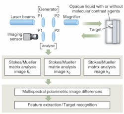
One of those techniques relies on the application of multispectral polarimetric techniques together with the use of unique molecular-contrast agents and biomolecular nanophotonic techniques. The key feature of this technique is the interrogation of targets or samples at different depths with polarized light at different wavelengths, forming multispectral Stokes-parameters-based images, such as degree-of-linear-polarization (DOLP) images.7, 8 Specifically, each light photon of a different wavelength will reach different depths; therefore, different-energy photons backscattered from those depths form spectral images. A subtraction of these spectral DOLP images gives rise to enhanced DOLP image differences due to the removal of the background surrounding the target, namely:
Here, S0, S1, and S2 are the Stokes parameters, estimated using the polarimetric measurement matrix method.9
These images can be further manipulated or combined to enhance the detection process. Experimentally, there are several approaches to measure the Stokes parameters utilizing the Mueller matrix formalism. In one study, the polarimetric arrangement of the Rotating Retarder Polarimeter Method was combined with the Polarimetric Measurement Matrix Method to obtain single DOLP images.10 In a typical example, DOLP images for a microsphere embedded in a scattering solution are acquired at 633 and 785 nm (see Fig. 2). In both cases, the target appears blurred and barely distinguishable in both images, although the image of the target acquired at 785 nm exhibits a slightly better contrast than the image at 633 nm. The DOLP subtraction image of the microsphere embedded in the same solution provides an enhanced image without distortion.
null
Efficient contrast agents
Further contrast enhancement of the target can be obtained by modulating the background of the target through doping with polar molecules and high-index-of-refraction molecules. This action leads to enhancement of the image contrast resolution (defined as the difference between the target signal and background signal over the arithmetic mean of the target signal and the background), as well as to reconfigurable, light-beam-steering capabilities, and increased magnification and depth of focus.
Besides the intrinsic potential of background doping for bulk imaging applications, this technique may lead to the development of effective cancer treatment methodologies by selectively doping areas surrounding tumor cells with dedicated high-index-of-refraction nanoparticles and polar molecules. These particles and molecules can generate pronounced resonance and local field effects, creating tumor-killing mechanisms similar to those observed using surface plasmonic nanostructures (such as gold nanoparticles or gold nanoshells).
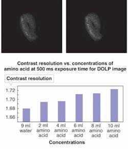
Alternatively, identifying the presence of precursor amino acids and enzymes in precancerous stages could lead to early cancer detection and treatment. The DOLP images of a test target consisting of a white twisted plastic wire immersed in water and aqueous concentrations of polar dopants were compared (see Fig. 3). A net improvement of the contrast resolution of the target results when the background of the target is modulated by introducing polar dopants. In this example, the amino acid L‑phenylalamine was used as the molecular dopant. A plot highlights the effect of amino-acid dopants on the image contrast as a function of increasing concentrations of the molecular dopant.
null
Future biophotonic devices
Another study looked at the potential for insulin molecules and other proteins-enzymes and dipolar ions alone or in conjunction with colloidal gold nanoparticles-to enhance the detection process of multispectral optical signals by providing unique signal characteristics, such as optical gain and light-shaping capabilities; on the other hand, the timing characteristics of such molecules, their stability, as well their long-term impact on the signal quality is under investigation.
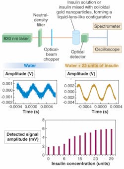
An insulin molecule contains a small number of hydrophilic groups on its surface, which explains its low solubility in water. Using a simple experimental setup, insulin molecules can be used to enhance and amplify incoming light signals (see Fig. 4). A beam of 830 nm photons passing through an aqueous solution of insulin exhibits a larger signal-to-noise ratio with respect to 830 nm photons detected after passing through the aqueous solution only. Larger signal-to-noise ratios are observed with increasing insulin concentrations; the overall enhancement of the signal characteristics can be plotted as detected light-photon amplitude (Vpp) versus aqueous insulin concentrations.
Further signal amplification and enhancement was observed by doping insulin molecules with colloidal gold nanoparticles. The overall improvement in signal quality could be attributed to the fact that acqueous insulin molecules would exhibit metamaterial-like characteristics manifested through pronounced self-focusing guiding and enhanced resonance effects, in combination with polarization mechanisms due to the rotation of permanent dipoles. These observations have significant implications for the search and identification of suitable optical bioinspired molecules and nanoparticles with stable optical characteristics, which would ultimately lead to the development of novel, scalable, highly reconfigurable biomolecular photonic materials, optical circuits and communication systems, tunable photonic nanocrystals, nanodevices, and therapeutic and high-performance imaging systems.
REFERENCES
1. G.C. Giakos, IEEE Trans. Instrumentation and Measurement55(6) 1904 (2006).
2. G.C. Giakos, Proc. IEEE Int’l. Workshop on Imaging Sys. and Tech. (IST), 150 (2007).
3. S.G. Demos et al., Optics Exp.7(1) 23 (July 2000).
4. M.A. Lecompte et al., Proc. SPIE2496, 180 (1995).
5. J.M. Bueno, J. Optics: Pure and Applied Optics, 470 (2001).
6. J.S. Baba et al., J. Biomedical Optics7(3) 341 (July 2002).
7. D.H. Goldstein and R.A. Chipman, Proc. Soc. Photo-Opt. Instrum. Eng.1745, 2007 (1992).
8. M.H. Smith, Applied Optics41(13) 2488 (May 2002).
9. D.B. Chenault and J.L. Pezzaniti, Proc. SPIE4133, 124 (2000).
10. R.M.A. Azzam, Optics Lett.2(6) 148 (1978).
GEORGE GIAKOS is director of and professor in the Imaging Technologies, Photonic Devices, and Molecular Nanophotonics Laboratories in the Electrical and Computer Engineering and Biomedical Engineering Department at The University of Akron, Akron, OH 44325-3904 and is also chairman of science and technology of Akron Scientific in Akron, OH; e-mail: [email protected]; www.uakron.edu.


