Advanced Imaging/Optical Design: Functional superresolution microscopy progresses
LAUREN ALVARENGA
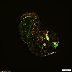
Traditional optical microscopy placed limits on what intracellular structures and activities could be imaged. Then superresolution techniques, led by software and equipment advances, made it possible to more clearly visualize the most minute details and processes that define life (see Fig. 1). Superresolution microscopy allows the fine mapping of cellular structures and specimens that are incompatible with electron or atomic force microscopy.
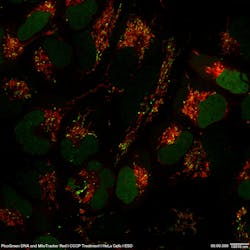
Further technology advances have enabled greater user-friendliness across a broader range of applications. Now, many of these techniques are meeting the challenges of speed and phototoxicity concerns, making them appropriate for live-cell imaging experiments. This latest generation of microscopy techniques is called functional superresolution (see Fig. 2). Because of their ability to meet the most exacting demands of biological imaging, functional superresolution techniques are preferred when superresolution is needed.Two functional super-res camps
The primary process of functional superresolution is the localization of diffraction-limited emitter coordinates (for example, single fluorescent molecules), fitting their point spread functions (PSF) and reconstructing localizations to create a final specimen image. There are two major categories in which the various techniques can be grouped: deterministic and stochastic.
Stochastic superresolution techniques focus the coordinates of single molecules through a series of fluorophore activations and deactivations. This process is repeated for thousands of frames as images are acquired point by point. These methods, including photoactivated localization microscopy (PALM) and stochastic optical reconstruction microscopy (STORM), offer high spatial resolution (20–50 nm), but have low temporal resolutions ranging from 30 seconds to 30 minutes, depending on the application, the sample, and the fluorochromes used.
Deterministic superresolution methods such as stimulated emission depletion (STED) exploit optical responses to excitation light to enhance resolution. STED uses two laser beams: one excites the sample and stimulates fluorescent emission, while the second is applied at the initial point of excitation and depletes all fluorescence except for that occurring in a small, subresolution volume of the sample. The imaging system then scans the sample, collecting image data coming from the excited fluorochromes at each scanned location.
Another deterministic superresolution technique, structured illumination microscopy (SIM), separates and reconstructs the Fourier transform of an image by shifting several frames in an image by some phase, making use of image information that is disregarded by widefield imaging approaches. SIM uses closely spaced repeating bands of light that interact with the specimen structure to produce moiré patterns and excite the sample. The results of the moiré patterns are processed via high-speed algorithms to yield higher-resolution images than would be achieved through traditional unstructured widefield or total internal reflection fluorescence microscopy. High-speed data processing algorithms enable the viewing of superresolution images in a display window to view live cells, various cell structures, or other molecules in real time.
Saturated structured illumination microscopy (SSIM) is a technique that introduces nonlinear fluorochrome responses to produce higher-order harmonics, further enhancing SIM’s potential.
Because SIM can provide as much as twice the native resolution of a widefield microscope, coupled with the advances in high-speed data processing algorithms that have removed the barriers of temporal resolution, real-time superresolution is now possible with confocal SIM for imaging rapid intracellular dynamics.
Enhanced resolving power—and more
Aside from enhanced resolving power, which is the predominant advantage of the technology, modern superresolution microscopy confers a variety of other benefits, including:
Observation at depth. With superresolution, researchers can clearly observe not only the surface of the sample, but also up to 100 µm deep within the sample. Some techniques have even reported producing an improvement in resolution up to an order of magnitude, allowing the study of subcellular architecture and dynamics at nanoscale. This is further enhanced through frequent improvements to objective lenses from commercial manufacturers.
Three-dimensional imaging. Higher temporal resolutions allow researchers to obtain detailed three-dimensional superresolution image data. This is done with certain techniques that obtain many images during time-lapse imaging that contain single molecule signatures, followed by fine mapping to render a superresolution image. In principle, this process is repeated for other z-planes to construct a three-dimensional image.
Ease of use. Through the combination of intrinsic optic sectioning, fast data acquisition, and dual-color superresolution, superresolution microscopy techniques deliver high-quality images quickly.Knowing the limitations
While superresolution offers a wealth of potential, there are some inherent drawbacks, most of which become exaggerated as resolution increases. For instance, superresolution techniques often require the use of complex hardware, special fluorophores, and imaging buffers, which may eliminate them from the consideration set of many traditional imaging experiments. Additional challenges include:
Point of diminishing returns. As superresolution systems image increasingly smaller details, the population of responding fluorochromes decreases, requiring the development of new fluorochromes with higher quantum yields. Depending on the technique used, the quality of superresolution images can also vary in detail and appearance.
Sample reliability. Live samples may be harmed or adversely affected by superresolution imaging because of high excitation intensity or extended exposure times. The stress placed on the specimen can also have implications for data reliability, which can potentially outweigh the benefits of higher resolution.
Optical aberration. Varying refractive indexes can affect the optical properties of light. To achieve the highest possible resolution, immersion oils can be properly matched to the refractive indices of both live and fixed samples.
Even with the restrictions and limitations of superresolution, researchers must consider the benefit of incredible accuracy, given the right imaging conditions, of imaging structures down to 10 nm.
Your needs, and the future
To determine if superresolution is the right method for your application, start by weighing the pros and cons—specimen integrity vs. an increase in resolution, system flexibility and speed, etc.
Also take time to review the specific needs of your study. Can you achieve your imaging goals by simply adjusting contrast? For instance, with confocal imaging, lowering the background intensity can improve fluorochrome visualization and enhance image analysis. In some cases, this enhanced contrast is enough to provide sufficient resolution. Or is what you’re trying to view so small that research requires a superresolution system?
Consider the validity of all solutions and compare superresolution techniques against other microscopy methods, including widefield and confocal imaging. Then, opt for whichever method produces the most usable fluorescence emission while also providing all necessary data.
Thanks to rapidly advancing technologies and growing software sophistication, commercial imaging instruments are able to offer flexible, multimodal systems capable of producing images beyond the diffraction limit, opening up a world of possibilities. These microscopes also offer a variety of user-friendly technologies, putting superresolution now within the reach of lab staff without extensive optical backgrounds. With this new level of enhanced resolving power, experiments are now taking us into uncharted territory.
Only time will tell the future of superresolution microscopy, but further advances will likely allow researchers to image even smaller structures for longer periods of time and image deeper into living tissue.
Lauren Alvarenga is a product manager at Olympus Corporation of the Americas – Scientific Solutions Group, Waltham, MA; e-mail: [email protected]; www.olympus-lifescience.com.


