Fluorescence Microscopy Part 2: Customizing Filters and Lenses
Customizing Fluorescence Microscopes
Customization of a fluorescence microscope involves fine-tuning its technical details to selectively utilize narrow bands of light for capturing high-resolution fluorescence images. Unlike standard brightfield or darkfield microscopes, fluorescence microscopes utilize specific filtering techniques to isolate the emitted light from fluorescent materials or fluorophores, which emit light of a longer wavelength when excited by specific excitation wavelengths.
The three critical filters in a fluorescence microscope are the excitation filter, dichroic filter, and emission filter. The excitation filter is placed in the illumination path and allows only the excitation range of the fluorophore under inspection to pass through while filtering out all other wavelengths from the light source. The dichroic filter, positioned at a 45° angle between the excitation and emission filters, reflects the excitation signal towards the specimen and transmits the emission signal towards the detector. Lastly, the emission filter, placed within the imaging path, allows only the emission range of the fluorophore to pass through, blocking the entire excitation range.
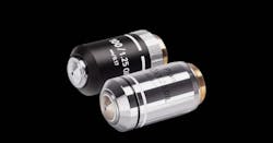
Lens customization is another aspect of enhancing the performance of a fluorescence microscope. There are several ways to customize lenses:
- Modifying the lens design: Optical design software is employed to optimize the shape and curvature of the lens to meet specific focal length, magnification, numerical aperture, distortion correction, or other requirements. This ensures the lens is tailored to the desired specifications.
- Special coatings: Coatings can be applied to the lens surface to minimize reflection and scattering, increase transmission in certain wavelength ranges, or enhance specific characteristics. For fluorescence microscopy, lenses may be coated to minimize the loss of fluorescence signal and provide high transmittance in the relevant wavelength range.
- Material modification: Different materials can be utilized to modify the lens’s performance and characteristics. For instance, materials with higher transmittance in specific wavelength ranges or greater durability under certain environmental conditions can be chosen.
- Special shapes and sizes: Custom lenses can be created in unique shapes and sizes, allowing for seamless integration into specific optics or equipment. Aspherical or asymmetric lenses may be used in specific applications.
Numerous advantages of customizing lenses for fluorescence microscopy:
- Optimal performance: Customized lenses are designed to meet specific requirements and applications, ensuring optimal optical performance and resolution.
- Suitability for special environments: Custom lenses can be selected to withstand harsh conditions like high temperatures, humidity, and vacuum, enhancing reliability and durability in specific applications.
- Enabling new technologies: Customized lenses may lead to the realization of new technologies and innovative applications, addressing unique research or industry needs and potentially yielding novel insights and results.
In summary, customization in fluorescence microscopy involves the precise selection and design of filters and lenses to efficiently capture and visualize fluorescence emissions. These customizations enable scientists and researchers to explore and understand biological processes at the single-molecule level and study live cells with high precision, contributing to advancements in various fields such as medicine, biology, and materials science.
Please contact us if you’d like to schedule a free consultation or request for a quote on your next project.
For other articles on Fluorescence Microscopy, please click here
Fluorescence Microscopy Part 1: Illuminating Samples for High-Resolution Imaging
