Live-Cell Imaging with Advanced Microscope Technology
Key Takeaways
- Live-cell imaging is revolutionizing cellular biology, offering insights into cellular behavior, developmental stages, and drug impact. This method visualizes individual or collective cell movements, morphological changes, and gene/protein expression under a microscope, all while keeping the cells alive.
- The significance of live-cell imaging is in its capacity to provide direct, real-time insights, which is particularly crucial in studies involving patient-derived samples.
- Various systems, which feature advanced technologies like Kohler illumination and AI-based sample detection, empower researchers to explore cancer cells, bone marrow, and fibroblasts. Avantier offers a wide range of custom optics and components according to the needs of customers.
- Live-cell imaging’s adaptability to different sample repositories contributes to its widespread adoption. It holds immense potential for advancing drug treatments and personalized medicine.
In the field of cellular biology, researchers are increasingly turning to the innovative technology of live-cell imaging to uncover the nuances of cellular behavior, developmental stages, and the impact of drug treatments.
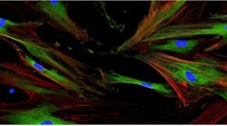
Live cell imaging is a method of visualizing and observing the movement and morphological changes of individual or collective cells under microscope, as well as the expression of genes and proteins, while keeping cells alive. Live cell imaging involves the observance of living cells for long periods of time, so it requires an incubator, a stage that can be maintained stably for long periods of time, and readjustment of focus.
A variety of biological phenomena can be observed through time-lapse observation using transparent images, such as dynamic cell movement during embryonic development, cell division, cell migration of cancer cells, and neurite extension of nerve cells. It is also used to observe the expression process of genes and proteins using fluorescent dye staining and protein staining, and to confirm changes in cells over time. This pioneering approach involves the utilization of specially configured microscopes, advanced illumination techniques, and analytical software to track and visualize cellular processes in real-time.
Diverging from traditional methods, the epi-oblique illumination technique brings the illumination process into the incubator.
The significance of live-cell imaging lies in its capacity to offer direct insights into cellular behavior, often in three-dimensional space. This becomes particularly crucial in studies involving patient-derived samples, such as the isolation of fibroblasts for skin research.
Different systems are employed for various levels of acquisition and analysis, featuring built-in Kohler illumination, motorized aberration correction, and automatic sample detection through pretrained artificial intelligence. This versatility encompasses various contrast imaging techniques, live-cell time-lapse imaging, and deconvolution, empowering researchers to delve deeper into the dynamics of cancer cells, bone marrow samples, and fibroblasts.
To cater to diverse needs in extended analyses, Avantier provides a range of solutions in custom optics and in custom lens assemblies.
The global market for live-cell imaging systems is anticipated to reach $4.18 billion by 2030, with a compound annual growth rate of 14.2% (Citation: https://www.strategicmarketresearch.com/market-report/live-cell-imaging-market). This growth is propelled by advancements in microscope technology and detector capabilities, rendering live-cell imaging increasingly feasible for drug discovery and cellular biology research. The adaptability of this technology to different sample repositories, including petri dishes, microplates, and flasks, contributes to its widespread adoption in laboratories worldwide.
In conclusion, live-cell imaging stands at the forefront of cellular biology, providing researchers with unprecedented insights into the dynamic world of cells. With its capability to picture cells under a microscope and to analyze cellular functions in real-time, this technology not only enhances our understanding of cellular behavior but also holds immense potential for advancing drug treatments and personalized medicine. As innovations like the epi-oblique illumination technique and cutting-edge imaging systems continue to emerge, the microscopic universe of live-cell imaging is poised for even more exciting discoveries in the future.

