SPECTROSCOPY/BIOSENSORS: SERS advance enables glucose detection, with implication for diabetics
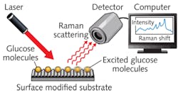
Glucose has very low Raman scattering efficiency, and existing substrates for surface-enhanced Raman spectroscopy (SERS) fail to amplify detection sensitivity to the level needed for finding concentrations typical in real samples. While the mechanism of SERS enhancement has not been well understood and is strongly dependent on the combination of surface and molecular target, a new substrate developed at the A*STAR-affiliated Singapore Bioimaging Consortium and Institute of Microelectronics provides enough of a boost to enable effective detection of glucose at physiological concentrations.1 The researchers hope that microsensors based on this resulting SERS platform may one day be implanted into patients for real-time glucose sensing—an advance that would dramatically improve the lives of people with diabetes.
Instead of the commonly used rough metal substrates, Malini Olivo and her co-workers etched silicon to form a well-defined pattern of nanogaps, and then coated the silicon with thin layers of silver and gold. Tests comparing the new substrate with commercial substrates for glucose detection showed an increase in sensitivity. “We were actually very surprised by our substrate’s high reproducibility,” says Olivo. “The best reproducibility reported previously for glucose was only about 10 percent. However, due to the special design and pattern of our substrate, we achieved reproducibility of about 3–4 percent, which is outstanding.” The nanogap substrate also provided good sensitivity for the detection of glucose in the physiologically important 0–25 millimolar range.
Now, the team is working on an analogous system for sensing proteins. “We would like to translate similar SERS substrate platforms to optical fibers in order to develop a minimally invasive in-vivo SERS platform for clinical diagnostics,” says Olivo.
1. U.S. Dinish et al., Biosensors and Bioelectronics 26, 1987–1992 (2011).
About the Author

Barbara Gefvert
Editor-in-Chief, BioOptics World (2008-2020)
Barbara G. Gefvert has been a science and technology editor and writer since 1987, and served as editor in chief on multiple publications, including Sensors magazine for nearly a decade.
