Leveraging oblique angle deposition (OAD) techniques used to grow nanostructured thin films, researchers at Pennsylvania State University (University Park, PA) have developed a new conformal-evaporated-film-by-rotation (CEFR) technique that is able to duplicate one of the most finely structured and complex biological systems: butterfly wings.
Butterfly wings are optically unique in that their overlapping roof-tile or scale structure (typically 200 to 600 scales/mm2) can present a range of high/low reflectivity values in a specific wavelength range, scatter light, or diffuse it over a broad wavelength range. This “physical” color presented to the observer has its origin in the fine wing structure. With practical application as optical filters for the development of miniature cameras for medical applications, replication of butterfly wings demands the fabrication of curved and complex nonplanar structures that to date have not been possible using traditional thermal or electron-beam evaporation, or sputtering techniques.
In OAD, a vapor flux is directed toward a substrate, with the trajectory of adatoms (atoms that lie on the surface) not parallel to the substrate normal. The arriving adatoms coalesce on the substrate to form thin films with an intrinsically anisotropic morphology, which can be tailored by altering the deposition conditions and using sequential substrate movements, thereby modulating the physical response characteristics of the films. The CEFR technique takes OAD one step further by spinning the biological specimen at a constant speed of about 0.5 rotations per second. The vapor flux is directed toward the sample at an angle of 85° to the substrate normal, creating a typical layer thickness of 0.5 µm. The entire process takes place in a noncorrosive environment that will not damage or distort the carbon/hydrogen/oxygen-based skeleton of the biological specimen. Finally, the original biotemplate is removed by plasma ashing, thus leading to self-standing biomimetic replicas.
The bulk material used for evaporation is a commercially available germanium antimonide selenide (GeSbSe) chalcogenide glass, chosen for its compatibility with the CEFR process and its interesting optical properties; namely, its large refractive index in the visible and infrared wavelength range, high transmittance in the infrared range, and the ability to modify its optical bandgap and refractive index by illumination. The bulk glass was thermally evaporated using a current of 95 A.
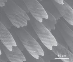
After evaporation, the visual appearance of the replicated butterfly wing was analyzed (see figure). The 200 x 50 µm scales have a complex surface structure with raised longitudinal ridges separated by a lattice structure with sub-micron features; in fact, this fine structure is what gives the butterfly wing its particular color.
Both the butterfly wing and the replicated structure compared favorably based on analysis of specular and scattered reflectance spectra, as well as direct and scattered transmittance spectra over the range 200 nm to 2 µm, indicating the high degree of fidelity for the replicated structure using the CEFR method. Slight differences in specular reflectance were assumed to be caused by the use of a different material (chalcogenide glass) to fabricate the replicated wing structure.
“CEFR is a powerful, versatile, and low-cost technique that allows the fabrication of replicas at the nanoscale made of very different materials, ranging from semiconductors to metals or insulators,” says Professor Raul J. Martin-Palmer, noting the technique can be used for the development of compound-eye-based miniature camers and optical sensors.
REFERENCE
- R.J. Martín-Palma et al., Appl. Phys. Lett. 93, 083901 (Aug. 25, 2008).
About the Author

Gail Overton
Senior Editor (2004-2020)
Gail has more than 30 years of engineering, marketing, product management, and editorial experience in the photonics and optical communications industry. Before joining the staff at Laser Focus World in 2004, she held many product management and product marketing roles in the fiber-optics industry, most notably at Hughes (El Segundo, CA), GTE Labs (Waltham, MA), Corning (Corning, NY), Photon Kinetics (Beaverton, OR), and Newport Corporation (Irvine, CA). During her marketing career, Gail published articles in WDM Solutions and Sensors magazine and traveled internationally to conduct product and sales training. Gail received her BS degree in physics, with an emphasis in optics, from San Diego State University in San Diego, CA in May 1986.
