Zapping cancer cells without collateral damage to healthy tissue. Delivering multiple drugs in controlled fashion to specific sites in the body. Diagnosing cancers and other diseases in their earliest stages. Imaging the interiors of cells in vivo. These are among the burgeoning possible uses of gold nanoparticles in biomedicine. Dozens of research teams around the globe now investigate the tiny particles’ potential as adjuncts to current methods of diagnosis and treatment.
Why do gold nanoparticles offer so many advantages? “It’s the fact that they’re nanometer-size systems that can get into the bloodstream and around cells,” explains Kimberly Hamad-Schifferli, assistant professor of biological and mechanical engineering at the Massachusetts Institute of Technology (MIT; Cambridge, MA). “They also have very unique optical properties. Many people are very interested in them because they change color when they bind to proteins. And the chemistry of the gold nanoparticles is very versatile; you can decorate them with biomolecules.” Equally important is the ability of research teams to create gold nanoparticles with several different shapes. Those physical differences give the particles versatility that is often lacking in other small-scale forms of diagnosis and treatment.
Physical versatility
Hamad-Schifferli and graduate student Andy Wijaya have used that physical versatility to design a system that can deliver multiple drugs into sites in the body and release them in a controlled way by firing infrared light from outside the body. The approach relies on two phenomena: Infrared light melts gold nanoparticles and causes them to release any items–such as drugs–attached to their surfaces. However, the infrared wavelength necessary to melt any particular nanoparticle depends on the particle’s shape.
The MIT researchers report in the journal ACS Nano that they created two shapes of gold nanoparticle. Wavelengths of 1,100 nanometers melt ‘nanobones,’ as the team calls them, while radiation at 800 nanometers melts ‘nanocapsules’. The team envisions creating up to four differently shaped particles to carry drugs into patients that physicians could release at appropriate times by using different infrared wavelengths. “With a lot of diseases, especially cancer and AIDS, you get a synergistic effect with more than one drug,” Hamad-Schifferli explains.
Two research teams have applied other shapes of gold nanoparticles to the destruction of tumors. In each, the particles first seek out cancer cells. Then, when exposed to near-infrared light, the particles destroy the cells by heating them. Because the heating is localized, it spares healthy cells. “This class of particle provides the most efficient method of specifically depositing energy in tumors,” explains MIT graduate student Geoffrey von Maltzahn.
The MIT group, headed by electrical engineering and computer science professor Sangeeta Bhatia, reports in the journal Cancer Research that it used gold nanorods to seek out and kill the tumors. The team also reports, in Advanced Materials, that it can enhance its nanorods’ ability to detect tumors by coating them with various light-scattering molecules.
Hollow nanospheres
The other team, headed by University of California, Santa Cruz professor of chemistry and biochemistry Jin Zhang, has used hollow gold nanospheres as a possible approach to treating malignant melanoma in a minimally invasive way; this devastating form of skin cancer kills more than 8,000 Americans annually. “Previously developed nanostructures such as nanorods were like chopsticks on the nanoscale. They can go through the cell membrane such only at certain angles,” Zhang told a meeting of the American Chemical Society. “Our spheres allow a smoother, more efficient flow through the membranes.”
Other types of gold nanoparticles have their own potential for diagnosis and cure. Scientists at Purdue University, for example, have developed a combination approach that can identify and reveal marker proteins on the surfaces of breast cancer cells. The team reported in the journal Angewandte Chemie that it has linked gold nanorods to particles of magnetic iron oxide. Optical microscopy identifies the luminescent nanorods and magnetic resonance imaging detects the magnetic particles at points too deep for luminescent detection. The probes also contain the Herceptin antibody that attaches to protein markers on cancer cells’ surfaces. “If we have a tumor, these probes should have the ability to latch onto it,” says associate professor of agricultural and biological engineering Joseph Irudayaraj, who headed the team. “The probes could carry drugs to the target, treating as well as revealing cancer cells.”
Star-shaped nanoparticles
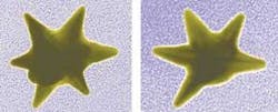
Bioengineers at Duke University (Durham, NC) have developed star-shaped gold nanoparticles. That shape has the advantage over other configurations of enhancing the amount of reflected light, thus enabling applications as contrast agents and labels for spotting pollutants and carcinogens. The technology relies on surface-enhanced Raman scattering (SERS). This occurs when light shines on a molecule; the molecule vibrates and scatters back light at its characteristic wavelength. Gold nanoparticles magnify the weak signals by a factor of more than a million. “We are trying to understand which types of nanostructures will give us the optimal signal so we can use them to monitor trace amounts of pollutants or detect diseases in their earliest stages,” says senior researcher Tuan Vo-Dinh. “This study is the first demonstration that these nanostars can enhance the effect of SERS to produce strong and unique signatures, like optical fingerprints.”Scientists at Singapore’s Institute of Bioengineering, meanwhile, recently reported in the Journal of the American Chemical Society that they have developed highly fluorescent gold nanoclusters for sub-cellular imaging. “Gold nanoclusters have promising characteristics for applications in vivo,” explains postdoctoral fellow Jianping Xie. “The red fluorescence of the nanoclusters enhances biomedical images of the body greatly, as there is reduced background fluorescence and better tissue penetration.” The team asserts that the process of synthesizing the gold nanoclusters can be easily scaled up for mass production.
Peter Gwynne | Freelance writer
Peter Gwynne is a freelance writer based in Massachusetts; e-mail: [email protected].

