Spectroscopic technique shows potential for early lung cancer detection
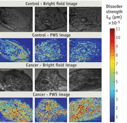
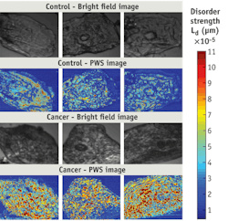
Partial wave spectroscopic (PWS) microscopy has now proven able to detect early signs of lung cancer by examining cheek cells in humans.1 While lung cancer survival rates are high when the disease is detected early, there are currently no recommended screening tests for large populations-so the disease is usually advanced by the time symptoms develop. The PWS method will distinguish those likely to benefit from more invasive and expensive tests from those who would not, according to Hemant K. Roy M.D., director of gastroenterology research at NorthShore University HealthSystem (Evanston, IL). Along with Vadim Backman, developer of PWS and professor of biomedical engineering at nearby Northwestern University, Roy earlier used PWS to assess the risk of colon and pancreatic cancers-also with promising results.
PWS can detect features as small as 20 nm in cells that appear normal using standard microscopy.
Roy and Backman conducted a study involving 135 participants: 63 smokers with lung cancer, control groups of smokers with and without chronic obstructive pulmonary disease (COPD), and 22 non-smokers. The test was equally sensitive to cancers of all stages, including early, curable cancers. Results were elevated (>50%) in patients with lung cancer compared to cancer-free smokers. A further assessment showed >80% accuracy in discriminating cancer patients from individuals in the three control groups. "The results are similar to other successful cancer screening techniques, such as the pap smear," Backman said, adding that the team hopes to improve early detection of other cancers, too. Backman and Roy think PWS has the potential to be used as a prescreening method, identifying patients at highest risk who are likely to benefit from more comprehensive testing such as bronchoscopy or low-dose CT scans. Next up: Additional large-scale validation trials.
1. H.K. Roy et al., Cancer Res. 70 (20): 7748-54 (2010)
More BioOptics World Current Issue Articles
More BioOptics World Archives Issue Articles

