Capsule endoscopy to gain remote-control capability
Capsule endoscopy—using a miniature camera system placed inside a small capsule that is swallowed and then transmits images while it journeys through the gastrointestinal tract—is not a new concept. Given Imaging (Yokneam, Israel) has been commercially producing a disposable capsule since 2001 for use in diagnosing intestinal and vascular disease (see Laser Focus World, January 2001, p. 83).
But researchers at City University of New York (CUNY) and City College of New York (CCNY) are taking the concept a few steps further, adding the ability to move the capsule via remote-controlled radio signals. The system may also one day be able to deliver remote-controllable treatment within the body, such as laser tissue removal or tissue welding.
Called the Compact Photonic Explorer (CPE), the capsule-based device is actually the brainchild of one of the scientist's children. A few years ago, after watching the movie Fantastic Voyage, 10-year-old Scott Alfano came up with the idea of miniaturizing a camera that could circulate through the body to perform diagnostic expeditions. His father, Robert Alfano, a CUNY distinguished professor of science and engineering at CCNY and the holder of 80 U.S. and foreign patents related to the science of optical diagnostics, consulted with CCNY electrical engineering professors to see if they could make the idea a reality.
Now the team has a U.S. patent for a "remote-controllable, microscale device for use in in-vivo medical diagnosis and/or treatment" and is collaborating with several other institutions to develop a spectroscopic-imaging version of the device. The four-year project, which combines engineering resources from Cornell University, SUNY Albany, Rochester Institute of Technology, Corning Rochester Photonics, Boston University, SUNY Binghamton, Rensselaer Polytechnic Institute, and the University of Rochester, is initially developing a CPE that can detect cancer and monitor body functions by combining imaging, polarization, spectroscopy, fluorescence, and biosensor technologies to relay information about the optical properties of a target object for real-time imaging and screening. Future CPEs will be designed to sense biological and chemical species, such as bacteria and pollutants, and monitor the health of compact structures and devices.
"One of the primary benefits of photonic imaging is that it can be conducted in a patient in vivo with little risk of injury," Alfano said. "Conventional imaging techniques, such as x-rays, expose patients to harmful substances—others, such as biopsy and histological evaluation, cannot be conducted in vivo. In addition, one of the current limitations of using photonics to image, examine, or treat tissue inside a patient is the difficulty of using optical fibers or endoscopes to access the tissue."
Multiwavelength imaging
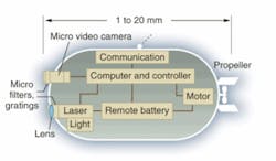
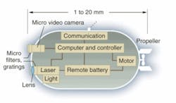
The CPE prototype measures 5 mm in length and comprises a transport capsule that contains imaging, data-collection, and data-transmission capabilities (see figure). It comprises a CMOS detector array, red, green, blue and near-IR light-emitting diodes (LEDs), radio-frequency (RF) transmitter, control electronics, and a miniature power source. The acquired images are transferred to a remote computer and receiving station by RF transmission. A multiwavelength imaging technique can detect an image in a cluttered background by comparing the differences in reflection and absorption properties between the target and the background under different wavelength illumination. Images of objects subjected to different colored illumination reflect and/or backscatter more light at nonabsorbing wavelengths.
Once the capsule is inside the body, an operator using radio controls and computer software can guide it to various locations, gathering two-dimensional image information and spectroscopic data along the way. Data is transmitted via video and radio signals to an external computer for use by an operator, who can make a diagnosis and instruct the device to render treatment to the affected area, such as the ablation of tissue using lasers or the binding of ruptured tissues with chemical glue, UV-cured epoxy materials, or photoionization techniques.
"The next step will be to use nanotechnology to make it even smaller," said Alfano, director of CCNY's Institute for Ultrafast Spectroscopy and Lasers, and principal investigator on the project. "The concept is to reduce the size of an endoscope from tabletop to watch to hand-held to pill to smaller, all using spectroscopy and LEDs. We're working from 280 nm to the infrared. Eventually, we want to do spectroscopy without moving parts, providing an endoscopy replacement that is less invasive, less costly, and more capable of scanning the entire digestive tract, from esophagus to colon."
In the detection and monitoring of cancer, future versions of the CPE will be able to evaluate subsurface skin lesions, according to Alfano. Analysis of images acquired with different-wavelength illumination can be used to determine the depth of skin lesions, an important factor in the treatment of melanoma.
"The objective is not just to shine light and get an image, but to send out colors, to explore inside the body and come up with answers, and then instruct the device to treat the problem," Alfano said.