Optogenetics: Optogenetics research drives new laser technologies
Optogenetics is proving to be an important photonics-based discipline impacting several areas of the life sciences. In neuroscience, in particular, it is enabling incredible advances in understanding brain function.
Currently, multiphoton excitation microscopy provides the only method of conducting optogenetic studies of live animal brains at single-neuron resolution. Here, ultrafast lasers may be used for any of several processes: to image the neural net anatomy, stimulate specific neurons, and to monitor resultant activity in interconnected neurons.
Early optogenetics experiments involved activating single neurons; however, the overarching trend today is to increase the number of neurons simultaneously triggered at high speed and with low temporal jitter. This need places unique demands on the output parameters of ultrafast lasers. Fortunately, ultrafast laser manufacturers are answering the call with important technological innovations to support ongoing advances in optogenetics experiments.
Manipulating cell membrane gradients
Much of biology involves the existence of electrochemical gradients across lipid membranes: difference in pH or concentration gradients in specific cations such as sodium, potassium, or calcium (Na+, K+, or Ca2+) or anions such as chlorine (Cl-). These gradients are controlled and maintained by proteins embedded in the membrane that act as either gates or pumps. Biologists have long studied these gradients using microscopic pipettes and electrodes in so-called patch clamp-type experiments.
More recently, confocal or multiphoton microscopy—combined with fluorescent probes and transgenic proteins that are sensitive to local electrochemical conditions (for example, calcium ion indicators)—have enabled these gradients to be monitored in a non-contact manner, in real time, with high spatial resolution in three dimensions (x, y, and z).
Pioneered by Karl Deisseroth's group at Stanford University (Stanford, CA) and Ed Boyden's group at the Massachusetts Institute of Technology (MIT; Cambridge, MA), among others, optogenetics provides a non-contact photonic method to actively manipulate these gradients using proteins acting as ion pumps that are turned on or off by light. The early work used the natural presence of proteins called opsins, but this soon led to the expression of light-activated proteins called channelrhodopsins (ChR1 and ChR2, for example) and then to other classes of light-sensitive genetically encoded tools to control local ionic concentrations—hence the term optogenetics.
By inserting genes for light-activated gates and pumps in subject organisms, optogenetics experiments run the gamut from studies on single muscle cells to macroscopic neurological effects. Dramatic examples of the latter involve illuminating part of a mouse/rat brain with light of the appropriate wavelength, causing the mouse to perform some involuntary action.1
Multiphoton excitation
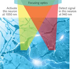
Optogenetics is ideal for brain function studies since signals are transmitted along neural axons in the form of a voltage spike (the action potential) across the axon membrane, mediated by rapid movement of cations across the membrane. In the outer layers of the mouse cortex, for example, spatially selective illumination using lasers and/or spatial light modulators (SLMs) enables target neurons to be stimulated or silenced. And in so-called "all optical physiology experiments," the related activity can be mapped as transient changes in fluorescent probes within the various interconnected neurons (see Fig. 1).2The most common fluorescent probe of this type—called genetically encoded calcium indicators (GECIs)—are the GCaMP family. Neurons expressing these probes show up brightly lit in the presence of Ca2+ activity and dark in its absence.
Because even the simplest part of the mammalian brain is a convoluted mass of countless interconnected neurons, an understating of brain activity requires experiments with high spatial resolution in three dimensions. Activity such as calcium ion concentration changes can be fluorescently monitored in a volume of live tissue by numerous wide-field and confocal laser microscope techniques that provide three-dimensional (3D) resolution. And while these techniques can also be used to map the structure of the brain volume under study, multiphoton excitation microscopy is often the preferred imaging method since it generally avoids the rapid photodamage that often occurs with confocal and live cells.
However, the only method that can be used to trigger activity at the single-neuron level with 3D resolution is multiphoton excitation. Here, two- (or sometimes three-) photon absorption has a highly nonlinear dependence on focused intensity, and thus only occurs at the focal plane of a tightly focused short-pulse (ultrafast) laser.
Longer wavelengths
Multiphoton excitation microscopy techniques and applications are traditionally supported by one-box titanium sapphire (Ti:sapphire) lasers optimized for these applications. For example, the Coherent Chameleon family provides a targeted combination of operational simplicity, short pulsewidths, and tunable wavelengths. The first multiphoton optogenetics experiments were successfully demonstrated with this type of laser, with numerous proof-of-technique experiments involving single-neuron stimulation.3
But it became apparent that many optogenetics experiments would soon outstrip the capabilities of Ti:sapphire-based sources in several areas-particularly power and wavelength range. As a result, laser manufacturers began to look at alternatives, including lasers based on ytterbium-doped gain materials.
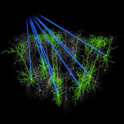
Because real brain activity involves the coordinated firing of myriad neurons, the optogenetics roadmap calls for experiments where hundreds and even thousands of neurons are excited and monitored at the same time. Ultimately, researchers would like to simultaneously survey as many as 10,000 neurons, which translates to a column of cortex measuring 250 × 250 µm with a depth up to 1 mm, spanning layers 1 through 6 of the mouse cortex (1 being the outermost layer).
It is well-proven that deep multiphoton excitation in live tissue benefits from longer wavelengths (at least 1 µm) to minimize scatter losses and other issues such as photodamage. And the challenges of separating the stimulation and detection/probing beams in optogenetics experiments are minimized if different wavelengths are used for these two processes.
Although a Ti:sapphire laser can be used together with a tunable optical parametric oscillator (OPO) for deep multiphoton excitation, other wavelength combinations are possible such as longer-wavelength optogenetics activators that are well developed and stable. An example is C1V1, which is effectively activated at approximately 1050 nm. Probing can then be accomplished at 920 nm with GCaMP, which has a low sensitivity to 1050 nm light. Unfortunately, only limited power is available at 1050 nm from even the best Ti:sapphire/OPO combinations.
In response, laser manufacturers developed a new type of fixed-wavelength laser based on cost-effective and reliable ytterbium-doped fiber technology. Technology once dictated that ultrafast fiber lasers based on ytterbium or erbium, as well as bulk lasers based on ytterbium, faced a tradeoff between power and pulse duration; that is, it has been nearly impossible to produce a sub-100 fs laser with power in excess of a few hundred milliwatts.
The Fidelity laser from Coherent has overcome this hurdle-delivering more than 2 W of power at 1055 nm. The simultaneous application of Fidelity and a Ti:sapphire laser to an all-optical physiology experiment at wavelengths of 1050 nm and 900–920 nm with approximately 2 W of power at each wavelength enables fast, parallel imaging, activation (or silencing), and Ca2+ signal detection (see Fig. 2). Researchers report that this laser combination can increase excitation from just a few neurons to several tens of neurons in a single experiment.4Even higher power
Despite the relatively recent release of these important fiber laser technologies, optogenetics researchers are already requesting even higher peak and average power in the 1050 nm region. Why the need for all this power in optogenetics? A satisfactory answer involves considering several factors.
First is the need to stimulate increasing numbers of neurons within a short time window. Membrane ion pumps operate on the timescales of a few milliseconds, and single-stage ion gates are even faster. Fast-scanning multiphoton microscopes with twin acousto-optic modulators (AOMs) are already proven for imaging large volumes and multiple regions of interest faster than video rates, so this type of microscope could easily direct a focused ultrafast beam at thousands of neurons in the requisite millisecond time window.
But the second part of the answer is that to fire or silence a neuron by photoactivation requires a relatively high fluence level, spread over the cell soma, even in the one-photon regime. Include the lower probability of two-photon absorption and the power requirement gets even larger.
Conversely, the low probability of two-photon absorption away from the focal plane means minimal photodamage, so the tissue can readily tolerate the long dwell times required. For example, in a 1997 paper from Schoenle and Hell, it was shown that 100 mW of power at 850 nm would not thermally damage a live sample even for an exposure of 1 s.5
With one-photon photoactivation, one solution is to enlarge the spot(s) to illuminate a much larger area of the membrane of the target neuron(s). But this is not really practical with two-photon excitation, where the already-low efficiency scales with the square of intensity.
At present, researchers are looking at several ways to best deploy the higher power they are pressing laser manufacturers to deliver. One way is to use "temporal focusing" of the laser in conjunction with an objective lens to produce a larger spot area (in the x-y plane), but without losing z-axis resolution (see Fig. 3).Temporal focusing was first demonstrated by Oron and Zhu et al. in 2005.6 Here, the laser beam is spread across an angular dispersive element such as a grating to create a chirped pulse. When refocused with a short-focal-length objective, the chirp disappears only when the beam is parallel again; that is, at the focal plane.
Spiral scanning is another solution being explored. Here, the focused beam is rapidly moved in an x-y spiral pattern to illuminate a membrane area much larger than the laser spot diameter using either twin galvanometer scanners or AOMs.3
Although the level of success in implementing these various techniques is difficult to predict, there is a clear and strong trajectory for using ultrafast lasers with higher powers in the 1050 nm window. Keeping pace with this demand for high powers has prompted laser manufacturers to look beyond traditional Ti:sapphire and to develop lasers using novel gain materials, most notably ytterbium-doped fibers and crystals. These new lasers provide the power necessary for today's cutting-edge experiments, together with the scalability to support anticipated future demand for single-neuron-resolution experiments in optogenetics.
REFERENCES
1. L. Fenno et al., Annu. Rev. Neurosci., 34, 389–412 and references therein (2011).
2. D. R. Hochbaum et al., Nat. Methods, 11, 8, 825–840 (2014).
3. J. Rickgauer and D. Tank, Proc. Nat. Acad. Sci., 106, 35, 15025–15030 (2009).
4. A. M. Packer et al., Nat. Methods, 12, 140–146 (2015).
5. A. Schoenle and S. W. Hell, Opt. Lett., 23, 5, 325–327 (1998).
6. D. Oron et al., Opt. Express, 13, 5, 1468–1476 (2005).
A tunable option
For some applications, it is highly advantageous to obtain two long wavelengths for all-optical physiology experiments from a single laser source. To address that need, laser manufacturers have developed a new generation of compact tunable lasers based on ytterbium (Yb) rather than titanium sapphire. An example of this type of laser is the Chameleon Discovery from Coherent, which uses nonlinear techniques to generate a tunable output from 680 to 1300 nm from a Yb-based, femtosecond pump source.
This tunable primary output has a pulsewidth of 100 fs with up to 1.4 W of output, well-suited for most multiphoton imaging applications. Plus, part of the direct Yb power (1.5 W) is available as a high-quality (m2 < 1.2) beam with fixed wavelength at 1040 nm, and short (160 fs) pulsewidth—ideal for long-wavelength photoactivators like C1V1. In addition, the availability of dual long-wavelength, phase-correlated pulses enables deeper imaging with techniques like coherent anti Stokes Raman scattering (CARS) and stimulated Raman scattering (SRS).
About the Author
Darryl McCoy
Director of Product Marketing and Senior Product Line Manager, Coherent
Darryl McCoy is Director of Product Marketing and Senior Product Line Manager at Coherent (Santa Clara, CA).
Marco Arrigoni
Marco Arrigoni is vice president of marketing at Light Conversion (Vilnius, Lithuania). He previously served as director of marketing at Coherent (Santa Clara, CA) from 2007 through 2023.
Nigel Gallaher
Product Manager, Coherent
Nigel Gallaher is a Product Manager at Coherent (Santa Clara, CA).


