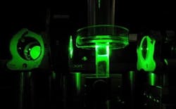
Using acousto-optics, Caltech researchers focus light inside biological tissue
Pasadena, CA--In a technique devised by researchers at the California Institute of Technology (Caltech), an ultrasound beam is used along with phase conjugation to create a high-power focal spot of light deep within a volume of light-scattering biological tissue.
In the technique, an ultrasound beam is tightly focused into scattering biological tissue so that it interacts with a beam of light passing through the tissue. The ultrasound acousto-optically frequency shifts the light, but only at the small ultrasound focus; that frequency-tagged scattered light can then be phase-conjugated after it exits the tissue and amplified, creating a time-reversed high-power beam that focuses tightly into the tissue at the same spot the ultrasound was focused.
Potential for surgery
While the previous limit for how deeply light could be focused into tissue was only about one millimeter, the Caltech technique reaches two and a half millimeters. In principle, the technique could focus light as much as a few inches into tissue. It could potentially allow doctors to perform surgery without having to cut through the patient's skin, or diagnose cancer by seeing tumors inside the body with a procedure that is as simple as an ultrasound.
Ying Min Wang, a Caltech graduate student in electrical engineering, and Benjamin Judkewitz, a postdoctoral scholar, are the lead authors on the paper, which was published in the June 26 issue of the journal Nature Communications.
The new work builds on a previous technique that Yang and his colleagues developed to see through a layer of scattering biological tissue, in which light scattered from tissue was used to record a hologram on a holographic plate. The recording contained all the information about how the light beam scattered; by playing the recording in reverse, the researchers could send light back through to the other side of the tissue, retracing its path to the original source.
To focus light into tissue, the researchers expanded on the recent work of Lihong Wang's group at Washington University in St. Louis (WUSTL), which had developed a method to focus light via ultrasound.
In both the WUSTL and Caltech experiments, ultrasound waves are focused into a small region inside a tissue sample, and frequency-shifting and phase conjugation then create a reversed beam. But the WUSTL experiment was limited, however, because only a very small amount of light could be focused. The Caltech engineers' new method, on the other hand, creates a higher-power beam.
The team demonstrated how the new method could be used with fluorescence imaging. The researchers embedded a patch of gel with a fluorescent pattern that spelled out "CIT" inside a tissue sample. They then scanned the sample with phase-conjugated focused light, which excited the fluorescent pattern, resulting in the glowing letters "CIT" emanating from inside the tissue. The team also demonstrated their technique by taking images of tumors tagged with fluorescent dyes.
With future improvements, the engineers say, they may be able to produce a focal depth of 10 cm—the depth limit of ultrasound—in a few years.
Sources: http://phys.org/news/2012-06-technique-focus-biological-tissue.html and http://www.nature.com

John Wallace | Senior Technical Editor (1998-2022)
John Wallace was with Laser Focus World for nearly 25 years, retiring in late June 2022. He obtained a bachelor's degree in mechanical engineering and physics at Rutgers University and a master's in optical engineering at the University of Rochester. Before becoming an editor, John worked as an engineer at RCA, Exxon, Eastman Kodak, and GCA Corporation.
