Large-field cryogenic objective mirror has nearly diffraction-limited performance

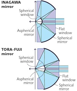
With a spatial precision of 1 nm, cryogenic fluorescence microscopy can image networks of biomolecules to a spatial precision of tens of nanometers or better. A group at Tokyo Institute of Technology (Tokyo, Japan) recently built such a microscope with subangstrom stability—to achieve this, the researchers designed and fabricated two types of monolithic cryogenic objective mirrors. The first, called the INAGAWA mirror, had a numerical aperture (NA) of 0.99, a very small image field of 1.5 µm radius, and operated at simultaneous wavelengths of 532 and 635 nm. The lens had an aspherical mirror, focal length of 2.11 mm, working distance of 1.0 mm, nearly diffraction-limited performance at a 1.7 K temperature, and an image stability of 0.05 nm over a 10-minute time span. But the small image field led to long acquisition times for sample-scanning of more than one hour. As a result, the group has now created a second cryogenic objective mirror, the TORA-FUJI, which has a slightly smaller NA of 0.93, but a much larger image field radius of 36 µm (large enough to image mammalian cells) and also exhibits nearly diffraction-limited performance at its illumination wavelength of 685 nm. Its focal length and working distance are 2.11 mm and 1.0 mm, respectively. (Both lenses operate while surrounded by liquid helium.)
The large field of view of the TORA-FUJI mirror was achieved by reducing the optical-design criterion “offense against the sine condition” (OSC), thus minimizing coma. The lens was tested using a single sCy7 dye molecule, which is much smaller than the diffraction-limited spot size, as a point source (no detection pinhole was used). The observed image patterns were consistent with a diffraction-limited point-spread function resulting from the annular lens aperture. As a bonus, the TORA-FUJI lens also provided nearly diffraction-limited performance at room temperature (296 K). The researchers next plan to use the objective for superresolution microscopy. Reference: M. Fujiwara et al., Appl. Phys. Lett., 115, 033701 (2019); https://doi.org/10.1063/1.5110546.

John Wallace | Senior Technical Editor (1998-2022)
John Wallace was with Laser Focus World for nearly 25 years, retiring in late June 2022. He obtained a bachelor's degree in mechanical engineering and physics at Rutgers University and a master's in optical engineering at the University of Rochester. Before becoming an editor, John worked as an engineer at RCA, Exxon, Eastman Kodak, and GCA Corporation.