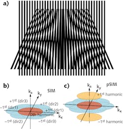
Polarization, a fundamental physical characteristic of light, has proven useful for various applications. In biology, polarization imaging reveals the orientation of targeted proteins by measuring the dipole orientation of fluorophores. Although superresolution polarization imaging has been used increasingly to study protein orientation, most such techniques are limited to labs with specialized expertise in these methods. Now, however, standard commercial structured illumination microscopy (SIM) systems can achieve superresolution polarization imaging, thanks to the work of researchers led by Peng Xi at Peking University and Qionghai Dai at Tsinghua University (both in Beijing, China).
In most SIM systems, high-frequency structured light is generated by the interference between two linearly polarized light sources (similar to Young's double slit interference); the superposition of structured light and the fine structure of the sample can produce Moire fringes. By detecting the Moire fringes at low frequency, the fine structure of the sample can be extracted.
Borrowing this principle, the researchers take the modulation of polarized light to dipoles as “angular structured light illumination,” therefore constructing a spatial-angular hyperspace and extracting the azimuth angle of fluorescent dipoles in a manner similar to SIM. The result is polarized structured illumination microscopy (pSIM), which, compared with other polarization imaging methods, offers three advantages simultaneously: high spatial resolution for imaging of ultrastructure, the ability to resolve dipole orientation of biomolecules, and the capture of intracellular dynamics.
Measuring dipole orientation
The pSIM image-reconstruction approach is based on the same dataset used by SIM, but pSIM extracts additional information. To achieve accuracy for dipole orientation measurement, the researchers determined polarization distortion by generating gratings using a custom setup with a spatial light modulator (SLM). They used a polarization beamsplitter to maintain linear polarization and high extinction in the diffracted laser beams, while rotating the beams’ polarization using a half-wave plate; a pair of dichroic mirrors placed on either side of a 4f optical system cancels the distortion. This enables an extinction ratio of greater than 10 before interference for each polarized beam.
To address the other factor that influences dipole orientation measurement, they calibrated nonuniform intensity using sparsely distributed 100 nm fluorescent beads to achieve fluorescence constancy under different polarizations until the resulting signal reported only intensity nonuniformity of the excitation over the entire field of view.
To prove compatibility with commercial SIM systems, the researchers demonstrated polarization superresolution imaging with 2D-SIM, 3D-SIM, and total internal reflection fluorescence (TIRF)-SIM imaging capabilities. To show its broad applicability for biological experiments, they also imaged a broad range of tissues and dynamics including λ-DNA, the actin filaments in BAPE cells and mouse kidney tissue, the interaction between actin and myosin, and green fluorescent protein (GFP)-stained microtubules in live U2OS cells (a bone-cancer line of cells). Furthermore, the team studied the membrane-associated periodic skeleton (MPS) in neurons. The high spatial resolution and accurate polarization detection capability of pSIM enabled the researchers to overturn the conventional concept of the “end-to-end” actin ring structure model, and revealed instead the “side-by-side” assembly model of actin ring in MPS.
Instant upgrade
Through their work to discover the polarization detection characteristics of SIM, the researchers realized that “the existing systems can be upgraded to polarization SIM system instantly, without any change in hardware,” according to Xi. “This enables life-science laboratories with SIM systems to directly access polarization SIM, which can greatly promote the research of polarization superresolution imaging.” Indeed, by combining the high spatial-temporal resolution of SIM with dipole orientation information, pSIM promises to help scientists answer a wide variety of biological questions.
REFERENCE
1. K. Zhanghao et al., Nat. Commun., 10, 4694 (2019).

Barbara Gefvert | Editor-in-Chief, BioOptics World (2008-2020)
Barbara G. Gefvert has been a science and technology editor and writer since 1987, and served as editor in chief on multiple publications, including Sensors magazine for nearly a decade.
