Biological tissue scatters light strongly, preventing imaging of subsurface features such as tumors. Optical coherence tomography (OCT) relies on broadband light and interferometry to overcome the effects of scattering of photons in tissue, allowing high-definition cross-sectional images to be made (see Laser Focus World, May 2000, p. 168). Acquiring data for an OCT image is done by scanning tissue in two dimensions: axially by means of a variable optical delay between the two interferometer paths, and transversely with a beam scanner. An endoscopic OCT system that can directly view internal organs needs to be compact and simple, whereas existing transverse scanning techniques tend to be neither.
Researchers from the University of Pittsburgh and Carnegie Mellon University (both of Pittsburgh, PA) have developed an OCT endoscope incorporating a microelectromechanical-system (MEMS) transverse-scanning mirror that is 1 mm square.1 All optics and mechanics fit within a 5-mm tube and could be made to fit into an even smaller tube. The MEMS mirror is made from a silicon wafer using a deep reactive-ion-etching complimentary metal-oxide semiconductor process. At one end of the mirror is a bimorph thermal actuator made of stacked aluminum and silicon dioxide thin films (top). An electrical resistor embedded in the bimorph mesh rapidly heats and cools the bimorph, pivoting the mirror. Nonlinear scanning effects can be reduced by careful design and by nonlinearly driving the resistor in a compensatory fashion, says Yingtian Pan, a University of Pittsburgh scientist. With a 165-Hz resonant frequency, the mirror scans a 2.9-mm transverse length at a 10-mm distance; an axial scanning range of 2.8 mm is achieved with a separate grating-lens-based optical delay line.
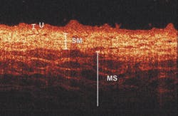
A two-dimensional endoscopic OCT image of a pig bladder wall in vivo (bottom) shows its important layers, including the urothelium (U), the submucosa (SM), and the muscularis (MS). A slight loss in image fidelity relative to a nonendoscopic OCT system resulted from coupling losses of greater than 3 dB in the endoscope. The MEMS-based OCT endoscope shows promise for early detection of bladder cancer, say the researchers.
REFERENCE
1. Y. Pan et al., Opt. Lett. (Dec. 15, 2001).

John Wallace | Senior Technical Editor (1998-2022)
John Wallace was with Laser Focus World for nearly 25 years, retiring in late June 2022. He obtained a bachelor's degree in mechanical engineering and physics at Rutgers University and a master's in optical engineering at the University of Rochester. Before becoming an editor, John worked as an engineer at RCA, Exxon, Eastman Kodak, and GCA Corporation.